Spermatogenesis and Spermiogenesis
The development of spermatozoa takes place in the male gonads called testes, which are paired in structure. In mammals, they are suspended in the scrotal sac, outside the abdominal cavity. The testes consisting of seminiferous tubules which remain enclosed within connective tissue and separated from each other by interstitial cells. The cell secretes male sex hormones called androgens. The seminiferous tubules are composed of both the germinal and somatic tissue. The germinal tissue lines the seminiferous tubules as a germinal epithelium which undergoes a process of spermatogenesis to form spermatozoa. The presence of somatic cells in the seminiferous tubules is a special feature of vertebrate testis called Sertoli cells.
Spermatogenesis is a continuous process of sperm formation. It is divided into two steps as under,
1. Formation of spermatids; 2. Spermiogenesis.
1. Formation of spermatids:
The cells of germinal epithelium when produced spermatozoa are called primordial germ cells. It undergoes the following phases,
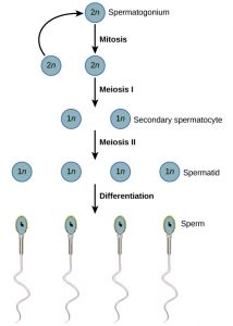
a. Phase of multiplication: The undifferentiated primordial germ cell contains a large-sized and chromatin-rich nucleus; it undergoes repeated mitotic cell division and becomes sperm mother cells or spermatogonia. Each cell is 2n (diploid) and enters into the phase of growth.
b. Phase of growth: During this phase, the spermatogonial cells accumulate a large amount of nutrition and chromatin material. Their volume becomes doubled and each cell is called the primary spermatocytes. It is 2n (diploid) in nature.
c. Phase of maturation: The primary spermatocytes enter the 1° meiotic or maturation division. During the division the nuclear DNA duplicate. The homologous chromosomes start pairing 1.e. form synapsis, each homologous chromosome splits longitudinally and by chiasma formation, the exchange of genetic material or crossing over takes place between the non-sister chromatids of homologous chromosomes. At the end of the first meiotic division, two secondary spermatocytes are formed. Each secondary spermatocyte is a haploid (n).
Each secondary spermatocyte undergoes a second meiotic or maturation division which is just like that of mitosis and produced 2 spermatids, which is haploid (n). Thus, at the end of meiotic division, a diploid spermatogonium produces four spermatids, which are non-functional male gamete, to become functional spermatozoa they have to undergo a process of differentiation or specialization, the spermiogenesis.

2. Spermiogenesis:
Differentiation of spermatids into the sperm is known as spermiogenesis. The sperm is a very active, motile cell, and this motility is provided by discarding superfluous material of developing sperm or spermatid. The (n) spermatid is a typical cell containing a nucleus and cytoplasm. The cytoplasm contains mitochondria, centrioles, and Golgi bodies. The two major parts of the spermatozoon are, the head and the tail (midpiece, principal piece, and end piece)become differentiated.
A] Formation of head:
The two major parts of the sperm head, the nucleus, and acrosome undergo the following changes to form the sperm head.
a) Changes in the nucleus: The nucleus of spermatids shrinks by losing much of water from the nuclear sap and assumes different shapes in different animals. The DNA becomes more concentrated and the chromatin material becomes closely packed. RNA, most of its proteins, and nucleolus are greatly reduced.
b) Acrosome formation: The acrosome of spermatozoan is derived from the Golgi bodies of spermatid. During acrosome formation, one or more vacuoles of Golgi bodies start enlarging and occupy the place between the tubules of Golgi bodies. Soon after a dense granule known as the proacrosomal granule develops inside the vacuole. The proacrosomal granule attaches with the anterior end of the nucleus and enlarges into the acrosome. The membranes of Golgi vacuoles form the double membrane sheath around the acrosome and form a cap-like structure of the spermatozoa. The rest of the Golgi bodies becomes reduced and discarded from the sperm as Golgi rest.
B] Formation of tail:
a) Centriole: The two centrioles of the spermatids become arranged one after the other just behind the nucleus. The anterior one is known as the proximal centriole and the posterior one is known as the distal centriole. The distal centriole changes into the basal bodies and gives rise to the axial filament of the sperm. The distal centriole and the basal part of the axial filament are present in the middle piece of the spermatozoa tail.
b) Mitochondria: Most of the mitochondria concentrate around the proximal part of the axial filament to form the middle piece of the spermatozoon tail. The tail of sperm comprises an axial filament which remains differentiated into a principal piece and end piece.
c) Change in the cytoplasm: Just prior to the termination of spermiogenesis, most of the cytoplasm is discarded from the sperm, but the plasma membrane forms an envelope around the head, middle piece, and main piece of the tail. The end piece of the tail remained naked and hence consist of only terminal parts of the axial filament.