Ultracentrifugation stands as one of the most powerful separation techniques in modern science, allowing researchers to isolate and study cellular components that would be impossible to separate by other means. This technique has revolutionized fields ranging from molecular biology to materials science by enabling the separation of particles based on their size, shape, and density – even down to the molecular level.
Principle of Ultracentrifugation
The fundamental principle behind ultracentrifugation is sedimentation. When a sample is rotated at extremely high speeds, particles experience a centrifugal force that causes them to move away from the axis of rotation. This force can be thousands or even millions of times greater than gravity, allowing for the separation of particles that would otherwise remain suspended in solution.
The rate at which a particle sediments depends on several factors, described by the Svedberg equation:
s = m(1-vρ) / f
Where:
- s = sedimentation coefficient (measured in Svedberg units, S)
- m = particle mass
- v = partial specific volume of the particle
- ρ = density of the solution
- f = frictional coefficient (related to shape and size)
Larger, denser particles sediment faster than smaller, less dense ones. By carefully controlling the centrifugal force and duration, scientists can achieve precise separation of biological molecules and cellular components.
Types of Ultracentrifugation
There are several different methods of ultracentrifugation, each suited to particular applications:
Differential Ultracentrifugation
This is the simplest form of ultracentrifugation, used to separate particles based on their sedimentation rate. The sample is centrifuged at progressively higher speeds, collecting the pellet after each run. This allows for the sequential isolation of cellular components based on their size and density.
Density Gradient Ultracentrifugation
This method uses a pre-formed density gradient medium to improve separation resolution. Two main types exist:
- Rate-zonal (size) separation: Samples are layered on top of a gradient and particles separate primarily based on their size and shape as they move through the gradient.
- Isopycnic (density) separation: Particles move until they reach a position where their density matches the surrounding medium. This creates distinct bands of separated components.
Analytical Ultracentrifugation
This specialized technique measures the properties of macromolecules in solution. It’s particularly useful for determining molecular weight, shape, and interactions between biomolecules.
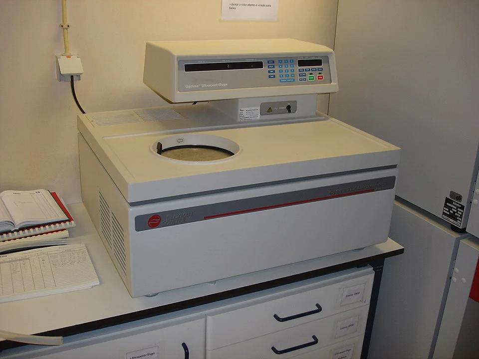
Parts of an Ultracentrifuge
An ultracentrifuge is an engineering marvel that must withstand enormous forces while maintaining precision. The main components include:
| Component | Function | Characteristics |
|---|---|---|
| Rotor | Holds the samples during centrifugation | Made of special titanium alloys for strength; multiple types available for different applications |
| Drive Motor | Provides rotational force | Capable of achieving speeds up to 150,000 RPM |
| Refrigeration System | Maintains temperature control | Prevents sample heating due to friction |
| Vacuum System | Reduces air friction | Maintains vacuum of about 10^-6 torr |
| Control System | Monitors and controls operation | Regulates speed, temperature, time, and safety features |
| Housing/Chamber | Contains all components | Heavy reinforced design for safety |
Types of Rotors
- Fixed-angle rotors: Hold tubes at a constant angle (typically 14-40 degrees). Best for pelleting applications and separating dense particles quickly.
- Swinging-bucket rotors: Tubes swing out horizontally during centrifugation. Ideal for density gradient separations with improved resolution.
- Vertical rotors: Hold tubes parallel to the axis of rotation. Provide the shortest path length for density gradient separations, reducing run times.
- Near-vertical rotors: Hold tubes at a slight angle from vertical (7-9 degrees). Combine advantages of vertical rotors with improved pelleting capabilities.
- Continuous flow rotors: Allow processing of large volumes by continuously pumping sample through the rotor.
Procedure for Ultracentrifugation
The ultracentrifugation process requires careful preparation and attention to detail:
Standard Procedure:
- Sample Preparation
- Prepare homogenized samples using appropriate buffers
- Filter or pre-clear samples if necessary to remove large debris
- Ensure proper ionic strength and pH for stability
- Rotor and Tube Selection
- Choose appropriate rotor type based on separation goals
- Select tubes compatible with the rotor and sample volume
- Verify tubes can withstand the planned centrifugal force
- Gradient Preparation (if applicable)
- Create density gradients using media like sucrose, CsCl, or Percoll
- Allow gradients to stabilize before use
- Carefully layer sample onto gradient
- Balancing
- Precisely balance opposing tubes (±0.01g)
- Use identical tubes, adapters, and caps
- Balance with matching solution when running fewer than maximum tubes
- Ultracentrifuge Operation
- Set refrigeration temperature (typically 4°C)
- Allow vacuum to establish before starting run
- Set acceleration/deceleration profiles
- Program speed and time parameters
- Monitor run for abnormalities
- Sample Recovery
- Use controlled deceleration to preserve separation
- For pelleting: carefully remove supernatant and resuspend pellet
- For gradients: collect fractions using specialized fraction collectors or by puncturing tube bottom
- Analysis
- Analyze fractions for presence of target molecules
- Use techniques like spectrophotometry, gel electrophoresis, enzymatic assays, etc.
Applications and Uses
Ultracentrifugation has diverse applications across scientific disciplines:
Biological Applications
- Subcellular Fractionation
- Isolation of organelles (mitochondria, nuclei, ribosomes, etc.)
- Separation of membrane components
- Isolation of protein complexes
- Virus Purification
- Concentration and purification of viral particles
- Separation of empty capsids from filled particles
- Determination of viral structure
- Protein Studies
- Determination of protein molecular weight
- Analysis of protein-protein interactions
- Characterization of oligomeric states
- Nucleic Acid Isolation
- Purification of DNA and RNA
- Separation of DNA fragments by size
- Isolation of plasmid DNA
Medical Applications
- Diagnostic Testing
- Separation of blood components
- Analysis of lipoprotein profiles
- Isolation of diagnostic markers
- Therapeutic Product Production
- Purification of vaccines
- Isolation of therapeutic proteins
- Preparation of blood products
Industrial and Research Applications
- Nanoparticle Characterization
- Size determination of engineered nanoparticles
- Purification of nanoparticle preparations
- Quality Control
- Testing homogeneity of pharmaceutical formulations
- Analysis of polymer characteristics
Comparison of Ultracentrifugation Techniques
| Technique | Principle | Resolution | Sample Capacity | Typical Applications |
|---|---|---|---|---|
| Differential | Sequential sedimentation at increasing speeds | Low to moderate | Large (10-100 mL) | Initial separation of subcellular components |
| Rate-zonal | Separation based on sedimentation velocity through gradient | High | Small to moderate (1-10 mL) | Separation of particles by size/shape |
| Isopycnic | Separation based on buoyant density | Very high | Small to moderate (1-10 mL) | Purification of nucleic acids, viruses |
| Analytical | Real-time analysis of sedimentation patterns | Very high | Very small (0.1-0.5 mL) | Characterization of biomolecular properties |
Safety Considerations
Ultracentrifugation involves considerable safety risks due to the extreme forces generated. Important safety measures include:
- Proper Rotor Maintenance
- Regular inspection for signs of stress or corrosion
- Adherence to manufacturer’s guidelines for maximum speed and lifetime
- Proper cleaning and storage to prevent corrosion
- Sample Containment
- Use of appropriate tubes rated for the intended speed
- Proper sealing to prevent aerosol generation
- Containment of hazardous materials
- Operational Safety
- Never attempt to open the centrifuge while running
- Ensure proper balancing before each run
- Follow laboratory biosafety protocols for hazardous samples
Frequently Asked Questions (FAQs)
Q: What’s the difference between a regular centrifuge and an ultracentrifuge? A: Ultracentrifuges operate at much higher speeds (up to 150,000 RPM vs typically less than 5,000 RPM for regular centrifuges) and generate forces in excess of 1,000,000 × g, whereas regular centrifuges typically generate forces below 5,000 × g. This allows ultracentrifuges to separate much smaller particles and molecules.
Q: How long does an ultracentrifugation run typically take? A: Run times vary greatly depending on the application, ranging from 30 minutes for some pelleting protocols to 48+ hours for equilibrium density gradient separations.
Q: What density gradient media are commonly used? A: Common media include sucrose (for rate-zonal separations), cesium chloride (for DNA/RNA), Percoll (for cells and organelles), and iodixanol (Optiprep, for biological materials sensitive to high salt).
Q: How do I know which rotor to use for my application? A: Rotor selection depends on your separation goals. Fixed-angle rotors are best for pelleting applications, swinging-bucket rotors for gradient separations requiring high resolution, and vertical rotors for faster isopycnic separations.
Q: What is the Svedberg unit? A: The Svedberg unit (S) is a unit of sedimentation coefficient, equivalent to 10^-13 seconds. It’s named after The Svedberg, who developed the analytical ultracentrifuge and received the Nobel Prize for his work in 1926.
Q: How can I determine protein molecular weight using ultracentrifugation? A: Molecular weight can be determined using analytical ultracentrifugation through either sedimentation velocity experiments (which measure how fast a molecule moves) or sedimentation equilibrium experiments (which measure the distribution of the molecule at equilibrium).
Q: How do I prevent my sample from heating during ultracentrifugation? A: Modern ultracentrifuges have refrigeration systems to maintain temperature. Additionally, the vacuum system reduces air friction that would otherwise generate heat. For sensitive samples, pre-cooling the rotor and samples is recommended.
References
- Cole JL, Lary JW, P Moody T, Laue TM. (2008). Analytical ultracentrifugation: sedimentation velocity and sedimentation equilibrium. Methods Cell Biol. 84:143-79. https://doi.org/10.1016/S0091-679X(07)84006-4
- Schuck P. (2013). Analytical ultracentrifugation as a tool for studying protein interactions. Biophysical Reviews. 5(2):159-171. https://doi.org/10.1007/s12551-013-0106-2
- Zhao H, Brautigam CA, Ghirlando R, Schuck P. (2013). Current methods in sedimentation velocity and sedimentation equilibrium analytical ultracentrifugation. Curr Protoc Protein Sci. Chapter 20:Unit20.12. https://doi.org/10.1002/0471140864.ps2012s71
- Rickwood D, Ford T, Steensgaard J. (1994). Centrifugation: Essential Data. Wiley. https://www.wiley.com/en-us/Centrifugation+Essential+Data-p-9780471942719
- Graham JM, Rickwood D. (2002). Subcellular Fractionation: A Practical Approach. Oxford University Press. https://global.oup.com/academic/product/subcellular-fractionation-9780199636730
- Beckman Coulter Life Sciences. (2020). Ultracentrifuge Applications and Methods. https://www.beckman.com/resources/technologies/centrifugation
- Griffith OM. (2010). Techniques of Preparative, Zonal, and Continuous Flow Ultracentrifugation. Beckman Coulter Inc. https://www.beckman.com/resources/fundamentals/centrifugation-guides