Introduction to Immunoglobulins
Immunoglobulins are glycoproteins produced by plasma cells (differentiated B cells) that function as antibodies in the immune response. They are found in blood, lymph, and other bodily fluids, where they identify and neutralize foreign substances, known as antigens, such as pathogens or toxins. The versatility of immunoglobulins lies in their ability to recognize a vast array of antigens due to their variable regions, making them indispensable for immune defense.
- Definition: Immunoglobulins are Y-shaped proteins that bind specifically to antigens, facilitating their elimination.
- Historical Context: The discovery of antibodies dates back to the late 19th century, with Emil von Behring and Shibasaburo Kitasato demonstrating their role in immunity, earning von Behring the first Nobel Prize in Physiology or Medicine in 1901.
- Role in Immunity: They are central to humoral immunity, targeting extracellular pathogens and toxins.
- Diversity: The immune system generates millions of unique antibodies to combat diverse antigens, achieved through genetic recombination and somatic hypermutation.
Structure of Immunoglobulins
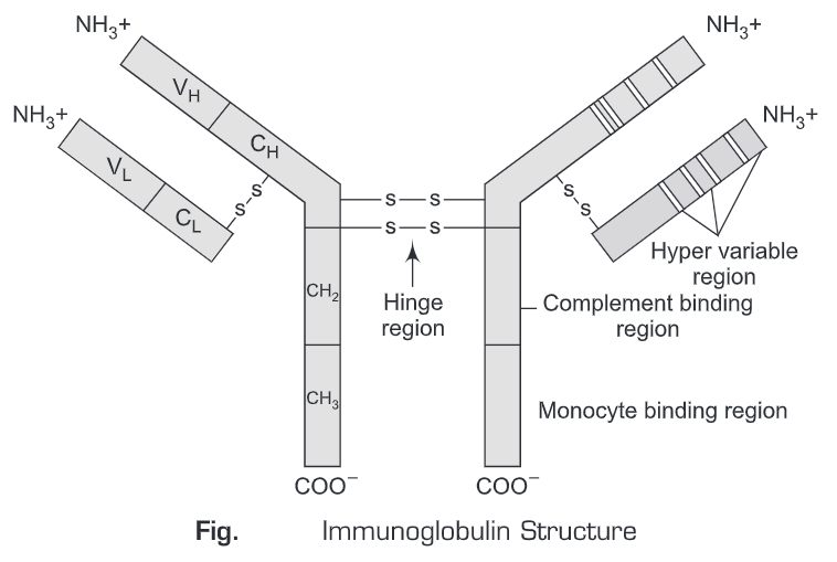
The structure of immunoglobulins is highly conserved across different classes, ensuring their ability to bind antigens and interact with immune cells. Their Y-shaped structure is composed of polypeptide chains linked by disulfide bonds, with distinct regions responsible for antigen binding and effector functions.
Basic Structural Components
Immunoglobulins consist of four polypeptide chains: two identical heavy chains and two identical light chains, connected by disulfide bonds.
- Heavy Chains:
- Larger polypeptide chains (~50-70 kDa).
- Determine the immunoglobulin class (e.g., gamma for IgG, mu for IgM).
- Contain a variable region (VH) and constant regions (CH1, CH2, CH3, or CH4 in some classes).
- Light Chains:
- Smaller polypeptide chains (~25 kDa).
- Two types: kappa (κ) and lambda (λ).
- Contain a variable region (VL) and a constant region (CL).
- Disulfide Bonds:
- Covalent bonds linking heavy and light chains, stabilizing the structure.
- Interchain disulfide bonds connect heavy chains to each other and to light chains.
- Hinge Region:
- Flexible region in heavy chains (except IgM and IgE) allowing the Y-shape to adjust for antigen binding.
- Contains disulfide bonds and is susceptible to enzymatic cleavage (e.g., by papain or pepsin).

| Component | Description | Role |
|---|---|---|
| Heavy Chain | Large polypeptide, class-specific | Defines immunoglobulin class, effector functions |
| Light Chain | Smaller polypeptide, κ or λ type | Contributes to antigen binding |
| Disulfide Bonds | Covalent bonds between chains | Structural stability |
| Hinge Region | Flexible segment in heavy chains | Allows conformational flexibility |
Domains and Regions
Each immunoglobulin chain is organized into domains, each approximately 110-120 amino acids long, formed by intrachain disulfide bonds creating a compact globular structure.
- Variable Domains (VH and VL):
- Located at the N-terminus of heavy and light chains.
- Contain hypervariable regions (complementarity-determining regions, CDRs) that form the antigen-binding site.
- Three CDRs per variable domain (CDR1, CDR2, CDR3) provide antigen specificity.
- Constant Domains (CH and CL):
- Form the bulk of the heavy and light chains.
- Responsible for effector functions (e.g., complement binding, Fc receptor interaction).
- Heavy chains have 3-4 constant domains (e.g., CH1-CH3 in IgG, CH1-CH4 in IgM).
- Fab and Fc Regions:
- Fab (Fragment antigen-binding): Contains VH, VL, CH1, and CL; responsible for antigen binding.
- Fc (Fragment crystallizable): Contains CH2 and CH3 (or CH4); mediates effector functions.
| Region | Domains Included | Function |
|---|---|---|
| Fab | VH, VL, CH1, CL | Antigen binding |
| Fc | CH2, CH3 (or CH4) | Effector functions (e.g., complement activation, Fc receptor binding) |
| Hinge | Flexible segment | Structural flexibility |
Antigen-Binding Sites
The antigen-binding site is formed by the variable regions of the heavy and light chains, specifically the CDRs.
- Complementarity-Determining Regions (CDRs):
- Three hypervariable loops in each variable domain.
- CDR3 is the most variable and critical for antigen specificity.
- Paratope:
- The specific region of the antibody that binds to an antigen’s epitope.
- Binding is non-covalent, involving hydrogen bonds, van der Waals forces, and hydrophobic interactions.
- Affinity and Avidity:
- Affinity: Strength of binding between a single paratope and epitope.
- Avidity: Overall binding strength, enhanced by multivalent interactions (e.g., IgM’s pentameric structure).
Classification of Immunoglobulins
Immunoglobulins are classified into five major classes (isotypes) based on the heavy chain type: IgG, IgA, IgM, IgD, and IgE. Each class has unique structural features and functions.
| Class | Heavy Chain | Structure | Molecular Weight | Serum Concentration | Key Locations |
|---|---|---|---|---|---|
| IgG | Gamma (γ) | Monomer | ~150 kDa | 9-13.5 mg/mL | Blood, tissues |
| IgA | Alpha (α) | Monomer/Dimer | ~160-320 kDa | 0.9-3.3 mg/mL | Mucosal surfaces, secretions |
| IgM | Mu (μ) | Pentamer/Monomer | ~900 kDa (pentamer) | 0.5-2 mg/mL | Blood, lymph |
| IgD | Delta (δ) | Monomer | ~180 kDa | 0.03-0.4 mg/mL | B cell surface |
| IgE | Epsilon (ε) | Monomer | ~190 kDa | 0.00005-0.0003 mg/mL | Mast cells, basophils |
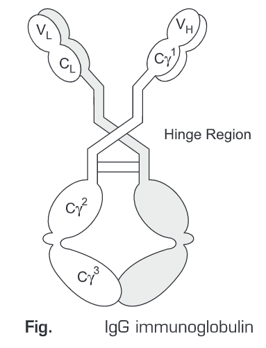
IgG
- Structure: Monomeric, with two Fab regions and one Fc region.
- Subclasses: Four in humans (IgG1, IgG2, IgG3, IgG4), differing in hinge region and effector functions.
- Functions:
- Neutralizes pathogens and toxins.
- Opsonizes pathogens for phagocytosis.
- Activates complement via the classical pathway.
- Mediates ADCC via Fc receptors on immune cells.
- Key Features:
- Most abundant immunoglobulin in serum (~80% of total Ig).
- Crosses the placenta, providing fetal immunity.
- Long half-life (21-28 days) due to FcRn receptor recycling.
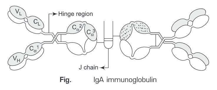
IgA
- Structure: Monomeric in serum, dimeric in secretions (secretory IgA, sIgA) with a secretory component and J chain.
- Subclasses: Two in humans (IgA1, IgA2).
- Functions:
- Provides mucosal immunity in secretions (e.g., saliva, tears, breast milk).
- Neutralizes pathogens at mucosal surfaces.
- Inhibits microbial adherence to epithelial cells.
- Key Features:
- Predominant in mucosal secretions.
- Secretory IgA is resistant to enzymatic degradation.
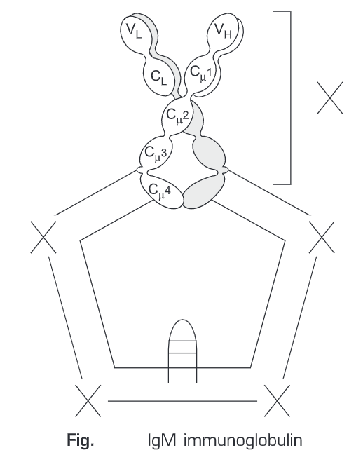
IgM
- Structure: Pentameric (five monomers linked by a J chain) or monomeric (on B cell surface).
- Functions:
- First antibody produced during an immune response.
- Highly effective at agglutination due to multiple binding sites.
- Activates complement efficiently.
- Key Features:
- Largest immunoglobulin due to pentameric structure.
- Short half-life (~5 days).
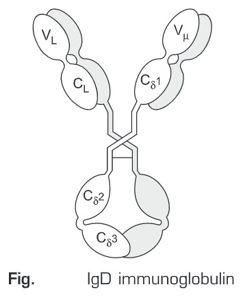
IgD
- Structure: Monomeric, primarily found on B cell surfaces.
- Functions:
- Acts as a B cell receptor, involved in B cell activation and differentiation.
- Minor role in serum immunity.
- Key Features:
- Low serum concentration.
- Role in immune regulation is still under investigation.
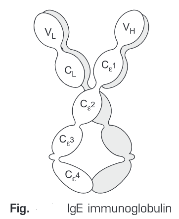
IgE
- Structure: Monomeric, binds to Fc receptors on mast cells and basophils.
- Functions:
- Mediates allergic responses and defense against parasites (e.g., helminths).
- Triggers histamine release in type I hypersensitivity reactions.
- Key Features:
- Lowest serum concentration.
- Long-lived when bound to mast cells.
Functions of Immunoglobulins
Immunoglobulins perform a variety of functions critical to immune defense, ranging from direct neutralization of pathogens to activation of other immune components.
Neutralization
- Mechanism: Antibodies bind to antigens (e.g., viral proteins, bacterial toxins), preventing them from interacting with host cells.
- Examples:
- IgG neutralizes viral particles, preventing cell entry.
- IgA blocks bacterial adhesion to mucosal surfaces.
- Significance: Prevents infection spread without direct destruction of the pathogen.
Opsonization
- Mechanism: Antibodies coat pathogens, marking them for phagocytosis by macrophages and neutrophils via Fc receptors.
- Key Players: IgG (especially IgG1 and IgG3) is highly effective due to strong Fc receptor binding.
- Significance: Enhances pathogen clearance by phagocytic cells.
Complement Activation
- Mechanism: Antibodies (primarily IgM and IgG) bind antigens, triggering the classical complement pathway, leading to pathogen lysis or opsonization.
- Process:
- Antibody-antigen complexes bind C1q, initiating the complement cascade.
- Results in membrane attack complex (MAC) formation or release of inflammatory mediators.
- Significance: Amplifies immune response and directly destroys pathogens.
Antibody-Dependent Cellular Cytotoxicity (ADCC)
- Mechanism: IgG binds to antigens on target cells (e.g., virus-infected cells), and Fc regions interact with NK cells, triggering cytotoxic granule release.
- Significance: Eliminates infected or abnormal cells.
Mucosal Immunity
- Mechanism: Secretory IgA prevents pathogen adhesion to mucosal surfaces and neutralizes toxins in secretions.
- Significance: First line of defense at mucosal barriers (e.g., gut, respiratory tract).
Allergic Responses
- Mechanism: IgE binds to allergens and Fc receptors on mast cells/basophils, triggering histamine release.
- Significance: Mediates type I hypersensitivity (e.g., allergies, anaphylaxis) but also protects against parasites.
| Function | Primary Immunoglobulin | Mechanism | Outcome |
|---|---|---|---|
| Neutralization | IgG, IgA | Blocks antigen activity | Prevents infection |
| Opsonization | IgG | Marks pathogens for phagocytosis | Enhanced clearance |
| Complement Activation | IgM, IgG | Triggers complement cascade | Pathogen lysis, inflammation |
| ADCC | IgG | NK cell-mediated cytotoxicity | Eliminates infected cells |
| Mucosal Immunity | IgA | Prevents adhesion at mucosal surfaces | Protects mucosal barriers |
| Allergic Responses | IgE | Histamine release | Allergic reactions, parasite defense |
Production and Regulation
Immunoglobulins are produced by B cells in a tightly regulated process involving antigen recognition, B cell activation, and differentiation into plasma cells.
- B Cell Activation:
- B cells recognize antigens via surface IgM or IgD (B cell receptors).
- Activation requires antigen binding and co-stimulatory signals (e.g., from T-helper cells).
- Class Switching:
- B cells switch from producing IgM to other isotypes (e.g., IgG, IgA) via somatic recombination.
- Driven by cytokines (e.g., IL-4 for IgE, TGF-β for IgA).
- Somatic Hypermutation:
- Introduces mutations in variable regions to increase antibody affinity.
- Occurs in germinal centers of lymph nodes.
- Plasma Cells:
- Differentiated B cells that secrete large quantities of antibodies.
- Short-lived (acute response) or long-lived (memory response).
- Memory B Cells:
- Provide long-term immunity by rapidly producing high-affinity antibodies upon re-exposure.
Clinical Significance
Immunoglobulins have significant applications in medicine, diagnostics, and therapeutics.
- Immunodeficiency Disorders:
- Conditions like X-linked agammaglobulinemia result in low immunoglobulin levels, increasing infection risk.
- Treated with immunoglobulin replacement therapy (IVIG).
- Autoimmune Diseases:
- Dysregulated antibodies (autoantibodies) attack self-tissues (e.g., rheumatoid arthritis, lupus).
- Vaccines:
- Stimulate antibody production for long-term immunity.
- Monoclonal Antibodies:
- Engineered antibodies used in cancer therapy (e.g., rituximab), autoimmune diseases, and infectious diseases.
- Diagnostic Tools:
- Antibodies used in ELISA, Western blot, and immunohistochemistry for detecting antigens.
FAQs
1. What are immunoglobulins?
Immunoglobulins, or antibodies, are Y-shaped proteins produced by B cells that recognize and neutralize antigens like pathogens or toxins.
2. How many types of immunoglobulins exist?
There are five main classes: IgG, IgA, IgM, IgD, and IgE, each with distinct structures and functions.
3. What is the difference between Fab and Fc regions?
The Fab region binds antigens, while the Fc region mediates effector functions like complement activation and phagocytosis.
4. Why is IgA important for mucosal immunity?
Secretory IgA prevents pathogen adhesion to mucosal surfaces, protecting areas like the gut and respiratory tract.
5. How do antibodies contribute to allergic reactions?
IgE binds allergens and triggers histamine release from mast cells, causing allergic symptoms.
6. What is the role of somatic hypermutation?
It introduces mutations in antibody variable regions to enhance antigen-binding affinity.
7. How are immunoglobulins used in medicine?
They are used in immunotherapy (e.g., IVIG, monoclonal antibodies) and diagnostics (e.g., ELISA).
References
- Janeway, C. A., Travers, P., Walport, M., & Shlomchik, M. J. (2001). Immunobiology: The Immune System in Health and Disease. Garland Science. Available at NCBI Bookshelf.
- Murphy, K., & Weaver, C. (2016). Janeway’s Immunobiology (9th ed.). Garland Science.
- Abbas, A. K., Lichtman, A. H., & Pillai, S. (2019). Cellular and Molecular Immunology (10th ed.). Elsevier.
- National Institute of Allergy and Infectious Diseases. (2023). Antibodies. Available at NIAID.
- World Health Organization. (2022). Immunization and Vaccines. Available at WHO.