
The model remains fundamental to cell biology, although modern research has revealed that the plasma membrane is more organized and complex than the original model suggested.
Fluid Mosaic Model Diagram
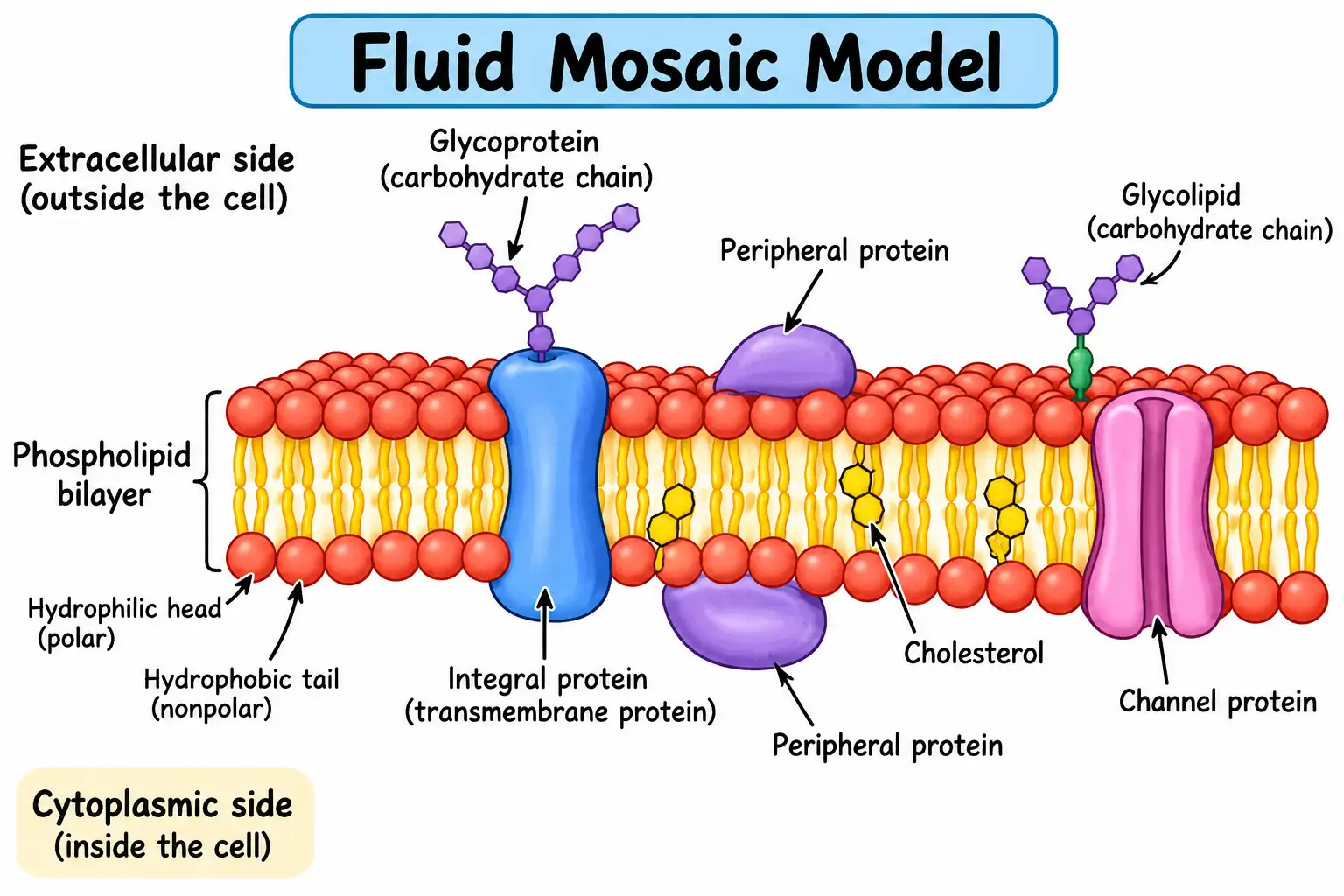
The diagram shows that the phospholipid bilayer forms the structural framework of the membrane, while proteins perform many specialized activities. Carbohydrate chains generally extend from the extracellular surface and participate in cell recognition and communication.
What Is the Fluid Mosaic Model?
The fluid mosaic model is a scientific explanation of the molecular organization of biological membranes. According to this model, the plasma membrane consists of a fluid phospholipid bilayer in which different proteins are embedded or associated with the membrane surface.
The term fluid refers to the ability of many membrane lipids and proteins to move laterally within the membrane. The term mosaic describes the varied arrangement of proteins and other molecules associated with the lipid bilayer.
A useful way to visualize the model is to imagine a flexible, two-dimensional sea of lipids containing different types of proteins. However, unlike objects floating freely in water, membrane proteins interact with surrounding lipids and may be anchored to other cellular structures.
Who Proposed the Fluid Mosaic Model?
The fluid mosaic model was proposed by S. J. Singer and G. L. Nicolson in 1972 in their research paper published in Science, titled The Fluid Mosaic Model of the Structure of Cell Membranes.
Their model replaced earlier interpretations that depicted the membrane as a relatively rigid structure with continuous protein layers coating the lipid bilayer.
The Singer–Nicolson model introduced a more realistic explanation of membrane organization, recognizing that proteins are distributed within and around a dynamic lipid bilayer.
Structure of the Fluid Mosaic Model
The plasma membrane consists of several interconnected components, each contributing to membrane stability, flexibility, and biological function.
1. Phospholipid Bilayer
The phospholipid bilayer is the basic structural framework of the cell membrane.
Each phospholipid molecule is amphipathic, meaning it contains two chemically different regions:
Hydrophilic head: A polar region that interacts with water.
Hydrophobic tails: Nonpolar fatty acid chains that avoid direct contact with water.
In an aqueous environment, phospholipids spontaneously organize into a bilayer. Their hydrophilic heads face the extracellular fluid and cytoplasm, while their hydrophobic tails point inward toward one another.
This arrangement forms a stable but flexible barrier, separating the internal environment of the cell from its surroundings.
The hydrophobic interior restricts the movement of ions and most polar molecules. Small nonpolar molecules, such as oxygen and carbon dioxide, can generally cross the bilayer through simple diffusion.
2. Integral Membrane Proteins
Integral membrane proteins are embedded within the lipid bilayer.
Some extend partially into the membrane, whereas others span the entire membrane and are known as transmembrane proteins.
These proteins perform several essential functions, including:
Transporting ions and molecules through channels and carriers.
Acting as receptors for hormones and other signaling molecules.
Participating in enzymatic reactions.
Connecting the cell membrane to the cytoskeleton or extracellular structures.
For example, potassium channels selectively allow potassium ions to pass through the membrane under appropriate conditions.
3. Peripheral Membrane Proteins
Peripheral proteins are loosely associated with the inner or outer membrane surface rather than embedded in the hydrophobic core.
They commonly interact with integral proteins or polar membrane components.
Their functions include providing structural support, participating in intracellular signaling, and helping organize the cytoskeleton.
For example, some peripheral proteins connect the plasma membrane with actin filaments, helping maintain cell shape and mechanical stability.
4. Cholesterol
Cholesterol is an important membrane component in animal cells.
It is positioned between phospholipid molecules, where it influences lipid packing and membrane fluidity.
At relatively high temperatures, cholesterol can restrict excessive phospholipid movement. At lower temperatures, it helps prevent phospholipids from packing too tightly.
In this way, cholesterol helps regulate membrane fluidity and permeability.
Important: Cholesterol is especially characteristic of animal cell membranes. Plant membranes contain other sterols, while most bacteria lack cholesterol and may use alternative membrane-stabilizing molecules.
5. Glycoproteins
Glycoproteins are membrane proteins with attached carbohydrate chains.
In the plasma membrane, these carbohydrate chains usually project toward the extracellular environment.
Glycoproteins participate in cell recognition, adhesion, receptor activity, and immune responses.
For example, certain cell-surface glycoproteins help immune cells distinguish between different cellular signals and interact with other cells.
6. Glycolipids
Glycolipids are lipids with attached carbohydrate groups, primarily located on the outer leaflet of the plasma membrane.
They contribute to the protective carbohydrate-rich surface known as the glycocalyx and are involved in cell recognition, communication, and membrane stability.
Some glycolipids also serve as specific binding sites for microbial toxins and other biological molecules.
Main Characteristics of the Fluid Mosaic Model
The fluid mosaic model explains several important properties of biological membranes.
| Characteristic | Explanation |
|---|---|
| Fluid nature | Many lipids and some proteins move laterally within the membrane. |
| Mosaic arrangement | Different proteins are distributed among membrane lipids. |
| Selective permeability | The membrane regulates the movement of substances. |
| Flexibility | The membrane can bend and change shape without losing continuity. |
| Membrane asymmetry | The inner and outer leaflets differ in lipid and protein composition. |
| Dynamic organization | Membrane components interact, reorganize, and form specialized regions. |
Why Is the Plasma Membrane Called Fluid?
The plasma membrane is described as fluid because its phospholipids are not held in permanently fixed positions.
They can move sideways within their own membrane leaflet. This lateral movement contributes to membrane flexibility and allows many components to reorganize.
The movement of a phospholipid from one leaflet to the other, known as flip-flop, is much less frequent without the assistance of specialized enzymes.
Membrane fluidity is influenced by temperature, cholesterol content, fatty acid saturation, and interactions between membrane components.
Why Is the Plasma Membrane Called Mosaic?
The term mosaic refers to the heterogeneous arrangement of proteins and other membrane-associated molecules.
Different proteins serve different roles, including transport, signaling, structural support, and enzymatic activity.
Their distribution creates a varied molecular pattern rather than a uniform coating of proteins.
Functions of the Fluid Mosaic Model
The fluid mosaic model explains how membrane structure supports several essential cellular functions.
1. Selective Transport
The plasma membrane controls which substances enter and leave the cell.
Small nonpolar molecules may pass directly through the lipid bilayer, whereas ions and most polar solutes require specific transport proteins.
Movement occurs through processes such as simple diffusion, facilitated diffusion, and active transport.
For example, the sodium–potassium pump uses energy from ATP to maintain important ion gradients across animal cell membranes.
2. Cell Signaling
Membrane receptor proteins detect signals from outside the cell.
When a signaling molecule binds to a suitable receptor, it can trigger intracellular processes that influence metabolism, growth, differentiation, or other cellular responses.
3. Cell Recognition
Surface glycoproteins and glycolipids help cells recognize one another.
A familiar example is the ABO blood group system, in which different carbohydrate structures on red blood cell surfaces determine blood group antigens.
4. Cell Adhesion and Structural Support
Membrane proteins help connect cells to neighboring cells and to the extracellular matrix.
They also interact with the cytoskeleton, providing mechanical support and helping maintain the shape of the cell.
5. Enzymatic Activity
Certain membrane proteins act as enzymes and catalyze biochemical reactions.
For example, membrane-associated enzymes are important in cellular energy metabolism and signal transduction.
6. Membrane Flexibility and Vesicle Formation
The flexible organization of membrane lipids enables processes such as endocytosis, exocytosis, and vesicle trafficking.
These processes allow cells to internalize substances, release secretory products, and transport materials between intracellular compartments.
Factors Affecting Plasma Membrane Fluidity
Membrane fluidity depends on the chemical composition and environmental conditions of the membrane.
| Factor | Effect on membrane fluidity |
|---|---|
| Temperature | Increasing temperature generally increases lipid mobility. |
| Unsaturated fatty acids | Double bonds usually reduce tight lipid packing and promote fluidity. |
| Saturated fatty acids | Straighter chains generally pack more tightly and reduce fluidity. |
| Cholesterol | Buffers changes in fluidity and influences membrane packing. |
| Fatty acid chain length | Shorter chains generally promote greater fluidity than longer chains. |
These effects depend on the membrane’s overall composition. Cells can adjust their lipid composition to maintain membrane function under changing environmental conditions.
Evidence Supporting the Fluid Mosaic Model
Several experimental observations support the dynamic organization of the plasma membrane.
Freeze-fracture electron microscopy: This technique revealed particles associated with membrane fracture faces, consistent with proteins embedded in lipid bilayers.
Cell fusion experiments: Experiments using differently labeled membrane proteins demonstrated that certain proteins can redistribute laterally after membranes fuse.
Fluorescence recovery after photobleaching (FRAP): In this technique, fluorescently labeled membrane molecules are photobleached in a small area. Recovery of fluorescence can provide information about the movement and mobility of membrane components.
Together, these approaches helped establish that biological membranes are dynamic rather than permanently rigid.
Limitations of the Fluid Mosaic Model
Although the Singer–Nicolson model remains a foundation of membrane biology, it does not completely describe the complexity of living cell membranes.
Modern studies have demonstrated that:
Membrane proteins do not always move freely; some are restricted by the cytoskeleton or other molecular interactions.
Membrane lipids and proteins may assemble into specialized domains.
Membranes are crowded with proteins, rather than containing widely separated protein molecules.
Membrane composition and organization differ between cell types and organelles.
Interactions with the extracellular matrix and neighboring cells influence membrane behavior.
Modern membrane models therefore retain the basic concept of a fluid lipid bilayer while recognizing membrane domains, molecular crowding, and regulated component mobility.
Difference Between the Fluid Mosaic Model and Sandwich Model
Before the fluid mosaic model, the Davson–Danielli model described the membrane as a lipid bilayer coated by protein layers.
| Feature | Sandwich model | Fluid mosaic model |
|---|---|---|
| Proposed by | Davson and Danielli | Singer and Nicolson |
| Year | 1935 | 1972 |
| Protein arrangement | Continuous layers covering lipid bilayer | Proteins embedded in or associated with lipid bilayer |
| Membrane fluidity | Not adequately represented | Dynamic lateral mobility |
| Current status | Historically important but superseded | Foundational model, subsequently refined |
Frequently Asked Questions (FAQs)
1. What is the fluid mosaic model in simple words?
The fluid mosaic model describes the cell membrane as a flexible phospholipid bilayer containing different proteins that carry out transport, signaling, and structural functions.
2. Who proposed the fluid mosaic model and in which year?
S. J. Singer and G. L. Nicolson proposed the fluid mosaic model in 1972.
3. Why is it called the fluid mosaic model?
It is called fluid because many lipids and proteins can move laterally, and mosaic because different proteins and molecules form a varied arrangement within the membrane.
4. What is the main component of the fluid mosaic model?
The phospholipid bilayer forms the basic structural framework, while membrane proteins perform many specialized functions.
5. What is the role of cholesterol in the plasma membrane?
Cholesterol helps regulate membrane fluidity, lipid packing, and permeability, particularly in animal cells.
6. Is the fluid mosaic model still accepted?
Yes. It remains the basic framework for understanding biological membranes, although it has been refined to account for specialized membrane domains and restricted protein movement.
7. What are the major functions of the plasma membrane?
The major functions include selective transport, cell signaling, cell recognition, cell adhesion, maintaining cellular boundaries, and supporting vesicle formation.
8. What is the difference between integral and peripheral proteins?
Integral proteins are embedded in the lipid bilayer, whereas peripheral proteins attach to the membrane surface, often through interactions with other proteins.
Conclusion
The fluid mosaic model of the plasma membrane provides a fundamental explanation of how lipids and proteins are organized in biological membranes. Introduced by Singer and Nicolson in 1972, it describes the membrane as a dynamic phospholipid bilayer containing a variety of proteins.
This molecular organization makes the plasma membrane flexible, selectively permeable, and capable of supporting essential cellular processes, including transport, signaling, recognition, and adhesion.
Although modern research has expanded the model by identifying specialized membrane regions and restrictions on molecular mobility, the fluid mosaic concept remains central to understanding cell membrane structure and function.
References
Singer, S. J., & Nicolson, G. L. (1972). The fluid mosaic model of the structure of cell membranes. Science, 175(4023), 720–731. Original research.
Cooper, G. M. The Cell: A Molecular Approach. NCBI Bookshelf. Cell Membranes.
Nicolson, G. L. (2014). The Fluid—Mosaic Model of Membrane Structure: Still relevant to understanding the structure, function and dynamics of biological membranes after more than 40 years. Updated model review.