Anatomy of Tongue
- The tongue, a muscular organ, is divided for descriptive purposes into the body, which lies relatively free in the oral cavity, and the base, which is fixed to the hyoid bone. The base spans the oral cavity and pharynx.
- The dorsum of the body possesses the median sulcus, a shallow groove superficially dividing the tongue longitudinally in the midline into right and left halves.
- The surface mucosa exhibits specialized zones demarcating the remnants of the embryologic origin of the tongue. The sulcus terminal is may be observed as a posteriorly directed, V-shaped shallow groove separating the anterior two-third (or body) from the posterior one-third (base) of the tongue. The terminal sulcus is the developmental dividing line.
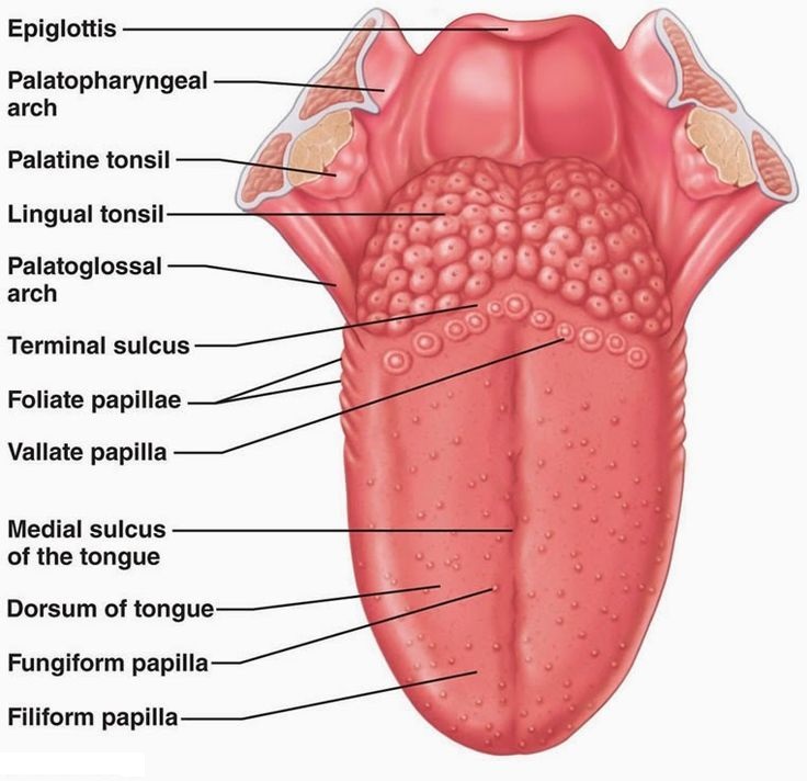
- That is, anything anterior to it is in the oral cavity, whereas anything posterior to it is in the pharynx. Lying alongside but anterior to the terminal sulcus is a row of 8 to 10 mushroom-shaped circumvallate papillae (vallate papillae).
- These structures possess taste buds and receive the ducts of the serous glands of von Ebner, one of the few named groups of minor accessory salivary glands.
- The remaining mucosal surface of the dorsum of the anterior two-thirds of the tongue possesses specialized projections, known as lingual papillae.
- The most numerous are the filiform papillae and, interspersed among them are the mushroom-shaped fungiform papillae; the former present a rough surface and they present no taste buds, whereas the latter display a few taste buds on their dorsal surface.
- On the posterolateral aspect of the anterior two thirds of the tongue are vertical furrows known as the foliate papillae; their taste buds degenerate after the first couple of years of life.
- Located in the midline, just posterior to the apex of the sulcus, is the foramen cecum, a shallow, pit-like depression that is a remnant of the developmental thyroglossal duct. The rest of the dorsal surface of the posterior one third of the tongue exhibits irregular bulges in its mucosa representing the lingual tonsils. The mucosa of the ventral surface of the tongue is smooth and without surface papillae.
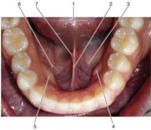
The medially placed lingual frenulum attaches the anterior two thirds of the tongue to the floor of the mouth. On either side of the frenulum, extending almost to the tip of the tongue, surface bulges may be observed representing the underlying glands of Blandin-Nuhn, another group of the named, minor accessory salivary glands. These glands are mixed, producing both serous and mucus saliva, which empty into the oral cavity via several minute pores.
- The bilateral deep lingual veins may be observed through the nearly transparent mucosa on either side of the frenulum coursing just deep to the mucosa along the tongue’s inferior surface from the tip to the deep regions in the floor of the mouth, where the vein disappears from view.
- Lateral to the vein is a fringed fold of mucous membrane, the plica fimbriata which often exhibits tissue tags from its free edge. Ducts of the glands of Blandin-Nuhn open into the oral cavity through the fringes of the plica fimbriata.
- Just above the floor of the mouth on either side of the lingual frenulum is an elevation of the mucous membrane (plica sublingualis) overlying the bulging sublingual glands.
- On closer examination one may observe several small openings along the surface of the plica sublingual is representing the small sublingual ducts (ducts of Rivinus).
- In addition, a large sublingual duct (duct of Bartholin) from the sublingual gland joins the submandibular duct (Wharton duct) just before its entry into the oral cavity for the delivery of saliva from the submandibular gland.
- The Wharton duct empties at the sublingual caruncula, an enlarged papilla adjacent to the lingual frenulum. Incisive glands, a small group of minor accessory salivary glands, may also be found on the floor of the oral cavity on either side of the lingual frenulum just posterior to the mandibular incisors.
References