Silkworm Diseases
Diseases:
- The silkworm, Bombyx mori that has been under domestication since time immemorial is prone to the attack of a number of diseases. Among the diseases pebrine, grasserie, flacherie and muscardine are important.
- They are all serious capable of destroying the entire stock (crop). Today, although no specific information on the loss due to various diseases is available, it is estimated that 30 to 40% of the total crop is lost due to diseases. Japan has reduced its loss to 10% in recent years.
Protozoan Disease
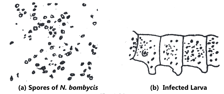
Pebrine: The Microsporidiosis of the silkworm is popularly known as ‘Pebrine’ all over the world. It is a French name; in Hindi it is called ‘Kata’. De Quartrifages (1860) gave the name pebrine to this disease because of the characteristic infection and appearance of dark pepper like black spots on the body of the infected larvae. It is first reported in France in 1845.
Pathogen: Pebrine is caused by a protozoan Nosema bombycis nageli belonging to the order Microsporidia. The infection occurs either via the mother moth or orally. The microspores found in the body of diseased silkworms are oval and refractile. Their longitudinal and transverse diameters are 2-3 µ and 1.5-2.0 µ respectively. Transovarian transmission of the parasite has been observed.
Symptoms: Infected larvae show black spots resembling pepper sprinkled all over the integument. The larva becomes milky white, unequal size, sluggishness, slow and irregular growths are the main symptoms. Infected pupae are heavier and the cocoons are flimsy. The moths that emerge from infected pupae are deformed by having small wing and distorted antennae. Further, the eggs are laid very irregularly. The silk is of inferior quality both in strength and uniformity.
Control:
- Use the disease-free female moths and eggs.
- The rearing house, equipments, eggs and workers were thoroughly sterilized with 2% formalin.
- Destruction of infected seeds and laying females.
Fungal Disease
Muscardine:
- The disease caused by fungal pathogens in insects is called muscardine disease. Depending upon the etiology and colour of the spore, different kinds of muscardine of silk worms are identified.
- They are white muscardine, green muscardine, yellow muscardine, red, purplish red, orange muscardine, aspergillosis, pencillosis etc.
The first three are of greater importance. However, aspergillosis is also important in recent times.
Following Table 1. shows the different kinds of muscardines of silkworms.
| Muscardine Type | Casual Organism |
| 1. White muscardine
| Beauveria bassiana (Bals)
|
| 2. Black muscardine
| B. brongniartti (Sacc)
|
| 3. Black muscardine
| Metarrhizium anisopliae
|
| 4. Green muscardine
| Spicaria prasina (Maublank Sawada)
|
| 5. Yellow muscardine
| Paecilomyces forinosus = Isaria farinosus (Dicks)
|
| 6. Red muscardine
| Sporosporella uvella (Krass)
|
| 7. Yellow red muscardine
| Paecilomyces fumosorosea
|
| 8. Purplish red muscardine
| Spicaria rubida
|
| 9. Orange muscardine
| Sterigmatocystis japonica
|
| 10. Aspergillosis/Brown muscardine
| Aspergillus flavus L., A. tamarii Kita
|
| 11. Pencillosis | Penicillium citrinum P. granulatum |
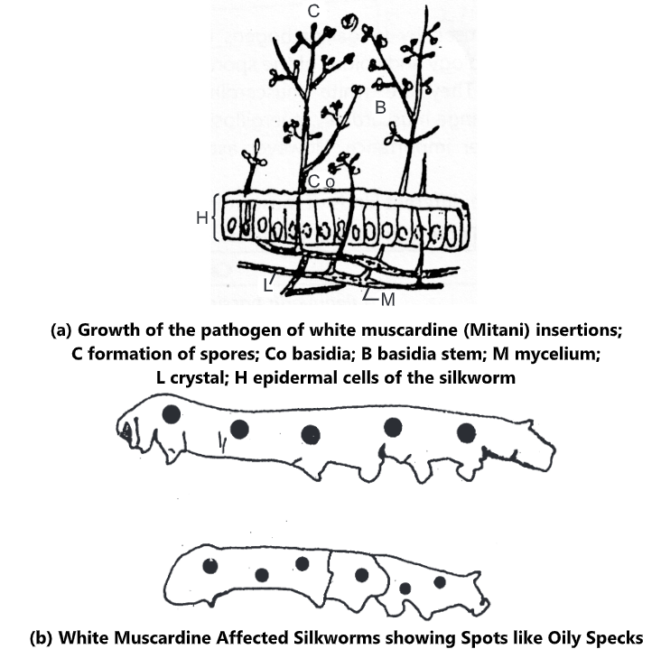
(a) White Muscardine: This disease is known by different names viz. Calcino, Sunnakaddi, Chitti and Chenakatu. It is first identified by Bassi in 1835.
Pathogen: Beauveria bassiana is causative organism. Its conidium is colourless, spherical or oval in shape, the size is 2.5-3.5-4.5 x 2.3-3.3-4.0 µ. Collectively they appear white in colour. The conidia germinate in about 6-8 hours at 26-28°C. The mycelia are colourless and branched.
Symptoms: In the early stage of the disease, the larvae lose appetite and become inactive. On progress, moist specks / oily stain appears on different parts of body. The larvae do experience diarrhoea and vomiting and consequently the body becomes limp losing its elasticity, around spiracles brown colour appears which later develops into black patch in 2-5 days of infection. After death, the body is soft initially and later becomes stiff and hard. This disease is very serious to aged silkworms.
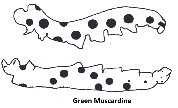
(b) Green Muscardine:
Pathogen: Spicaria prasina is causative organism; conidia are round or oval, light green in colour and measure 3.5 – 4.0-5.2×2.0-2.5-3.2 µ. Under favourable conditions (22-23°C) they expand, from an end. The germ tube penetrates into the body, ramifies and reaches the body fluid and organizes the hyphal body.
Symptoms: As the disease develops, the worms loss appetite and become inactive. Symptomatic specks of either round, oval or of irregular shape with the prominent periphery, clearly indicating the circumference, appear, which on coalescence form large specks. They appear dry and a little concave.
After death, the worms become stiff-hard and 2-3 days later the entire body becomes covered with mycelia and in 10-15 days later the mycelia bear fresh green conidia. This fungus also infects insects of various orders like Coleoptera, Lepidoptera and Hemiptera.
(c) Yellow Muscardine:
Pathogen: Paecilomyces farinose (= Isaria farinosa). The conidia are oval but rarely spherical or bell shaped collectively appear yellow and measuring 2.0-2.9-3.7 x 1.5-2.5-3.5 pin size. Under favourable conditions each gives out 1-2 germ tubes which form the ‘appressorium’ and later invade into host body.
Symptoms: In addition to becoming inactive, large specks appear around the spiracle and small specks appear over the body; with vomiting and diarrhoea. The body becomes hard and covered with yellow mycelia.
(d) Aspergillosis/Brown Muscardine:
This disease is caused by more than 10 species of the genus Aspergillus. Most of these fungi are distributed in the environment. They are saprophytic and become pathogenic only when conditions are favourable.
Pathogen: The important species are Aspergillus flavus Link, A. tamarii kite and A. oryzae. The conidiophores arise from mycelia and their tip is spheroidal or oval. The process of infection is almost similar to that of other muscardine fungi but the infection is localized and it does not extent to the body fluid.
Symptoms: The disease is very serious on chawki (I and II instar) worms. The infection is localised. Infected young worms become compact to lustrous and die soon. Mycelia emerge out from the spot of invasion and cause death. After death spore changes its colour with the age. Generally in the young worms, the body will not rot but in late age worms, the area which is not covered by mycelial mat will rot due to secondary infections. If the infection is by A. tamarii, initially it will be white and turn to greenish yellow, green, brown and finally to dark brown with large head like structures.
Bacterial and Viral Disease
Silkworm Flacherie: The term refers to flaccid condition of silkworm larvae suffering from dysentery. Flacherie is a ‘sub-chronic’ disease resulting primarily due to physiological disorders and secondarily by the infection of a bacterium.
(a) Viral Flacherie:
The viral flacherie is due to polyhedral and non-polyhedral viruses. Polyhedral viruses include cytoplasmic polyhedrosis virus, densonucleosis virus and kenchu virus diseases of silkworm.
(i) Infectious flacherie virus: It is caused by a virus of infectious flacherie I type.
Causal agent: Aizowa et. al. (1964) isolated spherical particles from diseased silkworms with a diameter of 30-32 nm and Kawase (1974) designated this as IFV.
Symptoms: Vomiting of digestive fluid, larval body looks transparent and sometimes body shrinkage is also observed. The infection is mainly through oral ingestion. Young worms are more susceptible to infection. The disease spreads through contaminated leaves.

(ii) Cytoplasmic Polyhedrosis: This disease is caused by a virus which infects the cytoplasm of the cell.
Pathogen and Infection: Polyhedra of the virus are found in the cytoplasm of cells. The size of polyhedra particle is 0.5 to 1.5 µ. It looks hexagonal under the microscope. The spread of disease occurs through faeces.
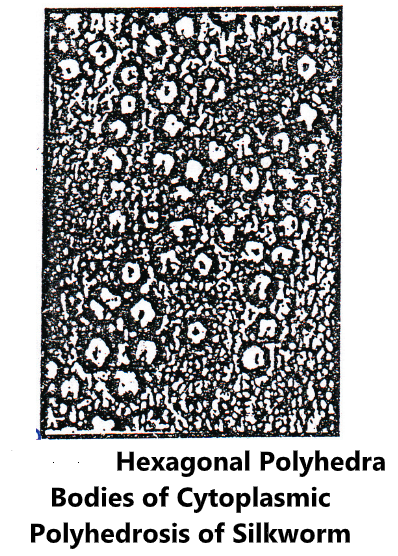
Symptoms: The infected worms show stunted growth leading to a prolonged larval life. On progress, the midgut becomes opaque and pale yellow, later it discharges whitish faeces and spoils the bed. Polyhedra are completely filled in cytoplasm of cylindrical and goblet cells are released into the alimentary canal as the cell is broken down. Consequently, polyhedra pass along with faeces, thus the spread occurs. Incubation period is about one week.
(iii) Kenchu disease of silkworm: This disease is referred as Kempu sappe, Kempunacchu and Nachu (Shyamala, 1978) and it appears to occur very frequently in Karnataka.
Pathogen: This is spherical or tetragonal, small, non-occluded virus of about 0.27 nm in diameter. Even single oral ingestion is enough to produce disease in all the stages of the insect.
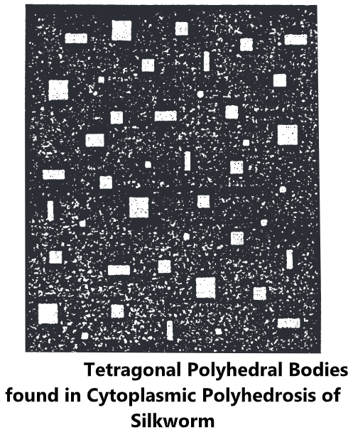
Symptoms: Immediately after feeding, the worms appear normal but after 1-2 hours, they show dullness, paleness and in some cases a disproportionately large head. The affected worms survive for several days but in a retarded state, dead larvae develop brownish patches. Sometimes mating of pupae is noticed when III, IV and V instar larvae are infected and they construct small and flimsy cocoons.
(b) Bacterial Flacherie:
This disease of worms caused by bacteria. It includes sotto, septicemia and Rangi (court) disease of silkworms. The bacterial diseases take a heavy toll of silkworm crop under high humidity. In Karnataka, the annual damage caused by this disease is 20-40%.
Pathogen and Infection: From the affected worms, many species of bacteria have been isolated. They are:
- Aerobacter cloacae
- Achromobacter superficialis
- Achromobacter delmarvae
- Staphylococcus albus
- Escherichia freundit
- Pseudomonas ovatis
- Pseudomonas boreopolis.
The spread and infection of disease is mainly by consumption of contaminated leaves. The dead larvae release pathogen to the surrounding and many are survived in the environment (air borne) within the rearing house.
Symptoms: In general, many species are involved in causing diseases which exhibit similar symptoms. The worm loses appetite and becomes sluggish. In the advanced stage, the larval body becomes very soft, discoloured and turns dark brown. Pulsation rate may increase rapidly and the worms wriggle due to pain. Later it vomits brownish fluid and passes very soft excreta which sticks to the rearing bed. Shadding of skin may not be proper during moulting.
(i) Sotto disease: The disease is caused by Bacillus thuringiensis Var. Sotto. Worms are killed by the toxin produced by the pathogen. Toxin is a rhombic crystal and is released out of the cell as the bacteria collapse. Pathogenicity is lost within 3 hours when in contact with formalin (3%) and within 5 minutes if boiled.
Symptoms: In the beginning, the worm loses appetite, becomes inactive, shows combination of flacherie symptoms and finally die. In the advanced stage, the silkworm loses appetite suddenly, writhes, affected with convulsions and dies in a short period. The victim turns gradually to brown, blackish brown or black and begins to rot.
(ii) Septicemia: The disease is caused when bacilli similar to those of sotto bacillus, or to colitis germs or B. prodigiosus etc., infected the worms.
Symptoms: The septicemia condition of the body results due to multiplication of a certain Bacillus in the body fluid. The worm loses appetite and becomes inactive. At times it vomits fluids, the abdominal legs loose gripping power and finally dies. After the death rot begins quickly. The invasion is mainly through wound on the skin and in some cases through the alimentary canal.
Viral Disease
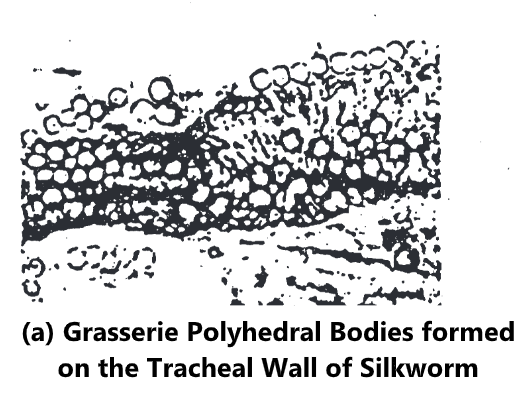
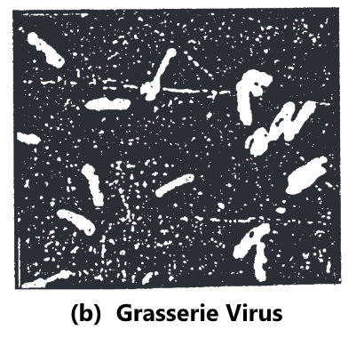
(a) Nuclear Polyhedrosis Virus (NPV):
In Karnataka, it is called as haluhula or haluthonde, in Italy as Giallume and in France as grasserie or Jaundice.
Causal Agent and Infection: The infection is mainly through mouth. After ingestion of the virus, the protein coat is dissolved in the gut releasing the viral rods. These rods themselves attack midgut cells and the infectious sub-units are set free which multiply in different cells and also enter the nucleus (NPV) of the susceptible cells and multiply. Small spherical bodies appear and on maturity form rods. Bundles of such rods are surrounded by proteins and they crystallize to form the polyhedra.
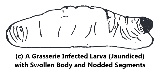
Symptoms: About a week after infection inter-segmental membranes are swollen and the larvae appear to be under stress. Haemolymph becomes turbid, skin loses its tension, becomes fragile and ruptures easily, releasing the milky haemolymph. The dead larvae are soft and flabby. Young ones loose coordination in movement and crawl about in circles and also fail to moult and die soon.
The skin of diseased pupae is fragile and disintegrates easily on handling and occasionally black marking appears on the body at the time of death. The disease mainly affects IV and V instar larvae. The environmental factors and quality of leaves also influence the incidence of the disease.