Cells of the Immune System are “stem cell, T cell, B cell, NK cell, Macrophages, Dendric cell”.
White Blood Cells
- Leukocytes/White Blood Cells/WBCs (5000 to 9000 per mm)
- Leukocytes or white blood cells are defensive cells that are important to both adaptive and innate host defenses.
- White blood cells (WBCs), also called leukocytes, are the cells of the immune system that are involved in defending or protecting the host body against infections and foreign invaders.
- Leukocytes are the largest cells of the blood although they are the least numerous blood cells. These cells are phagocytic, live for about three to four days in the average human body, and are found throughout the body.
- An increase in the number of leukocytes due to infection or allergies over the upper limits is called leukocytosis/leukemia.
- Some viral and rickettsial infections may cause a decrease in the leukocyte count called leucopenia.
- Types of leukocytes – Leukocytes can be classified either morphologically or functionally as Granulocytes and Agranulocytes.
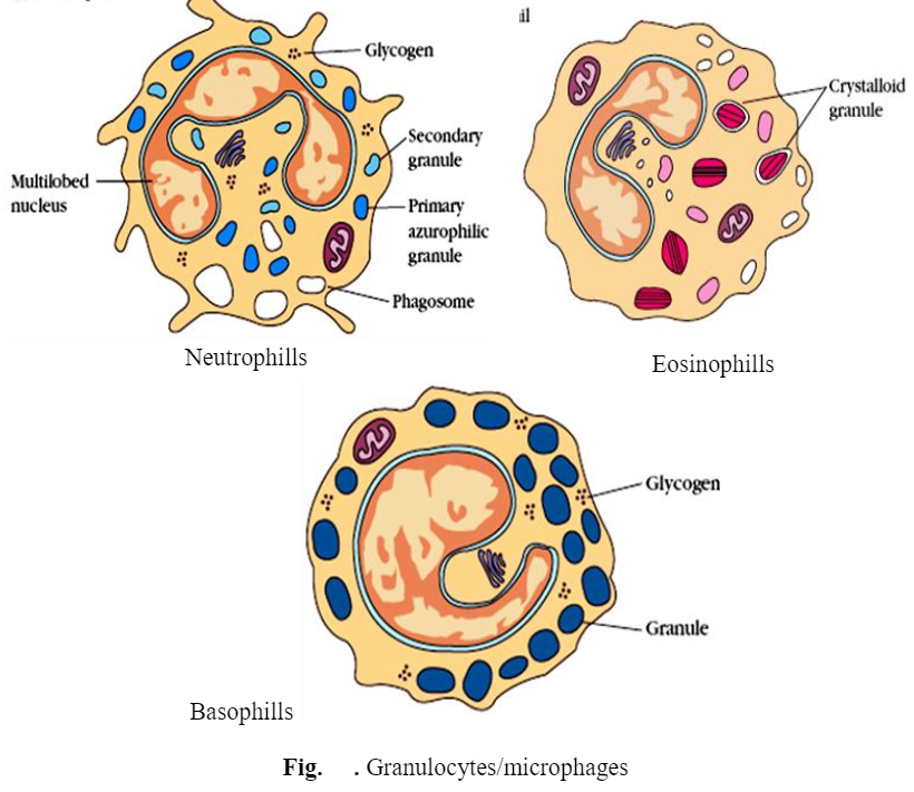
Granulocytes/Microphages
The granulocytes have granular cytoplasm and an irregularly shaped, labeled nucleus. Granulocytes can be distinguished from one another by the shape of their cell nuclei and by their staining reactions with specific dyes. Granulocytes include three types of cells —
a). Neutrophils, b). Eosinophils and c). Basophils.
a) Neutrophils (60% to 70% of total leukocytes) –
- The neutrophils are also commonly called Polymorph Nuclear Leukocytes (PMN), or Polymorphs.
- The term polymorphonuclear refers to the fact that the nuclei of neutrophils contain two to five lobes.
- The granules of neutrophils are stained red and blue with a mixture of acidic and basic dyes. Neutrophils are highly phagocytic and motile.
- Neutrophils guard the infection these cells are phagocytic and respond quickly wherever tissue injury has occurred. The cytoplasm may look transparent because of fine granules that are pale lilac.
- Neutrophils are active in phagocytosing bacteria and are present in large amounts in the pus of wounds. These cells die after having phagocytosed a few pathogens. The life span of a circulating human neutrophil is about 5.4 days.
b) Eosinophils (2-4 % of total leukocytes) –
- In general, their nucleus is bi-lobed. The cytoplasm is full of granules that assume a characteristic pink-orange color with eosin stain.
- Although eosinophils are physically too small to ingest and destroy helminths, they can attach to the outer surface of the parasites and discharge peroxide ions that destroy them.
- The numbers of eosinophils increase significantly during parasitic (Hookworm) infections and hypersensitivity reactions. This increase in the number of eosinophils is known as eosinophilia.
c) Basophils (2-4 % of total leukocytes) –
Basophils appear blue in color when stained by using the basic dye e.g. methylene blue. The nucleus is bi- or tri-lobed, but it is hard to see because of the number of coarse granules that hide it. They are characterized by their large blue granules.
 Mast cells
 Mast cells
- Mast cells occur in connective tissue and alongside blood vessels. They release histamine and are associated with allergies.
- Mast cells are generally quite similar to basophils in terms of morphology, receptors expressed, and function.
- They are not however homogeneous and there are two types of mast cells that exhibit some critical physiological and morphological differences.
- One population is found in the mucosa of the lungs and intestine and the other in the connective tissues near blood vessels.
- The differences in these cells are the number and size of granules, the density of receptors expressed, pharmacological properties, life-span, and response to drugs. This property of response to drugs can bear significantly on potential control and treatment of allergies.
Dendritic cells
- The dendritic cells are the antigen-presenting cells (APCs) found in the skin (Langerhan’s cells), thymus (medulla), lymph nodes (cortex and paracortex), spleen, and other secondary lymphoid organs.
- Dendritic cells are large, adherent motile cells. They possess numerous and prominent pseudopodia that represent approximately 1% of the cells in the tissues in which they reside.
- Langerhans cells with characteristic Birbeck granules (clubbed shaped) can migrate to the lymph nodes following binding of antigen and present the antigen to T cells in the paracortex and these follicular cells are also seen forming cellular interconnections in the cortical B cell areas.
Function of granulocytes
i) Phagocytosis – Microphages are the phagocytic cells responsible for the phagocytosis of certain parasites through the production of toxic proteins.
ii) Allergic reaction – During certain allergic conditions and parasitic infections, the numbers of eosinophils can increase dramatically. They can engulf and remove immune complexes and can inactivate certain allergic mediators.
iii) Damage to the antigen – When the parasite enters the body, eosinophils are activated through the complement system resulting in the release of major basic proteins and eosinophilic cationic protein which damages the outer layer of parasites.
iv) Maintenance of homeostasis – Leukocytes participate in maintaining homeostasis by 1) removing undesirable materials from the internal environment and ii) destroying noncontributing cells for transformed cells.
v) Detoxification – Eeosinophils may also detoxify foreign substances and help turn off inflammatory reactions. Eosinophils are phagocytic and have the ability to leave the blood.
vi) As anticoagulant – Basophils release heparin as an anticoagulant substance.
Agranulocytes
Agranulocytes are cells that lack the granular cytoplasm and have round nuclei. They include the following types of cells.
Monocytes
- Monocytes (3-7% of total leucocytes) are generally the largest cells of all WBCs. They lack the granules in their cytoplasm and are not actively phagocytic until they leave circulating blood and enter the body tissues and at last mature into the macrophages.
- Monocytes have many fine vacuoles containing digestive enzymes. They have a kidney-shaped nucleus and are typically agranulated.
- As the monocytes mature. their nucleus becomes oval or kidney-shaped having fine wrinkles. Monocytes enter the tissues and develop into macrophages. Monocytes are the third most common in the circulation (3-7%).
- Once monocytes move from the bloodstream out into the body tissues, they undergo changes (differentiate) allowing phagocytosis, and are then known as macrophages.
- Some leukocytes migrate into the tissues of the body to take up a permanent residence at that location rather than remaining in the blood. Often these cells have specific names depending upon which tissue they settle in, such as fixed macrophages in the liver, where they are known as Kupffer cells.

Functions of monocytes/macrophages
i) Phagocytosis – The primary function of macrophages is to phagocytize the Ag. When Ag succeeds in penetrating the skin or mucous membrane, local macrophages accumulate around the invaders and the Ag is phagocytized inside a vacuole called a phagosome. The membrane of the phagosome and lysosome fuses to form a phagolysosome. Lysosomal enzymes like acid proteases, RNAase, DNAase, phosphatase, and lipase digest the particle. However, some microbes like S. typhi, M tuberculosis, and Brucella abortus can resist digestion by macrophage. Monocytes functions as “vacuum cleaner”, and are much longer lived
ii) Ag processing and presentation – Macrophages trap the Ag and provide it in optimum concentration and an effective manner to the TH in such a way that it is easily recognized by T cells. Provision of optimum concentration of Ag is necessary, because a very low concentration of Ag may not be an antigenic and a very high concentration of Ag may be tolerogenic. In both cases, it will not induce the immune response.
iii) T cell activation – Macrophage secretes a number of biologically active substances such as interleukine 1 and leukocytes activating factor (LAF) which induces the synthesis of interleukin 2 by T cells. interleukine 2 facilitates the activation of T cells for cell-mediated immune response.
iv) Antitumor and graft rejection – Macrophages when stimulated by cytophilic antibodies and lymphokines, become armed. Such armed macrophages are capable of Ag-specific cytotoxicity which play important role in antitumor and graft rejection activities.
v) Energy cells –The recognition of Ag by T cells releases macrophage activating factors that differentiate the macrophage into Energy cells. Energy cells contain more lysosomes and are capable of vigorous phagocytosis.
vi) Prevention of infection – The vital role of macrophages is to prevent the infection. Deficiency of phagocytosis causes granulomatous disease, in which the patient succumbs to recurrent bacterial infection even though the T and B cell functions remain normal.
Lymphocyte
It Contains (20-40% of total leucocytes), It is a mononuclear leukocyte that mediates humoral or cell-mediated immunity. A lymphocyte is a white blood cell involved in specific immune responses. Lymphocytes are derived from lymphoid stem cells located in the bone marrow. Lymphocytes are the central cells of the immune system they are the immunological attributes of diversity and specificity of memory of self and non-self recognition. The lymphocytes constitute 20-40% of the body’s WBCs and 99% of the cells in the lymph. These lymphocytes continually circulate in the blood and lymph and they are capable of migrating into tissue spaces and in the lymphoid organs. Lymphocytes are not phagocytic but play a key role in specific immunity.
Types of lymphocytes – Lymphocytes include – B-cells, T cells, and NK cells
B-cells
- B-cells were first demonstrated for a special lymphatic gland of chickens called the bursa of fabrics the site for their maturation in birds.
- A lymphocyte that matures in the bone marrow and expresses membrane-bound antibodies is called B cells.
- After interacting with antigens these B-cells are differentiated into antibody-secreting plasma cells and memory cells.
- B-cell develops from stem cells located in the bone marrow in adults and the liver of a fetus B-cells probably stay in the bone marrow and mature there and after maturation, the mature B-cells migrate to the lymphoid organs such as the lymph nodes and spleen.
- This B lymphocyte-derived its letter designation from its site of maturation in the bursa of Fabricius in birds and this name turned out to be apt.
- B-lymphocyte is a white blood cell that gives rise to plasma cells and antibodies. Small spherical cells with a uniformly dark blue, rounded nucleus surrounded by a thin fringe clear pale blue cytoplasm.
- Humoral (Ab-mediated) immune response is carried out by Antibodies (Abs) which are produced by B-cells. The process of Ab formation (production) starts when B-cells are exposed to free, or extracellular antigens (Ags).
T-Cells
T-lymphocytes originate from stem cells in adult bone marrow or the fetal liver and these cells pass through the thymus and emerge as mature T-cells. T-cells are small, spherical cells with a uniformly dark blue, rounded nucleus surrounded by a thin fringe of clear pale blue cytoplasm. T-cell is one of the major types of lymphocyte T-cell develops from a stem cell processed in the thymus gland and this is responsible for the cell-mediated.
Functional types of T-cells
Regulatory cells — Regulatory T cells are of two types i) TH (Helper) and ii) TS (suppressor).
i) TH-cells — T Helper cells- TH cells play a central role in the immune response. The major function of TH cells is to stimulate other cells of the immune system to fight off intruders. TH cells activate cytotoxic T cells and other helper T cells, necessary for B-cell activation by T-dependent antigens. Helper T (TH) cells play a central role in the immune response. Their major function is to stimulate other cells of the immune systems to fight off introducers. For example, they activate cytotoxic T cells and other helper T cells. Some other TH cells also help B cells respond to antigens.
ii) TS (Suppressor T cells) – Suppressor T (Ts) cells are generally thought to be T cells that regulate the immune response by turning it off when an antigen is no longer present. Ts cells are also important in the maintenance of self-tolerance, preventing self-directed T cells that avoid clonal detection in the thymus from mounting immune responses. Some immunologists consider it likely that Ts are not a distinct population but present suppressive activity by the TH and Tc cell populations; however, Ts cells usually carry CD8 receptors, whereas most TH cells carry CD4 receptors.
T suppresser cells regulate immune response and help to maintain immunological tolerance. TS cells are also important in the maintenance of self-tolerance preventing self-directed T cells which avoids clonal deletion in the thymus from mounting immune response. Some immunologists consider it likely that Ts cells are not a distinct population but represent suppressive activity by the TH and Tc cell populations. However, Ts cells usually curry CD8 receptors and most TH cells carry (DH receptors).
- Effector T cells — Effector T cells include Tc cells DTH, MLR.
i) Tc cells – Activated cytotoxic T (Tc) cells destroy target cells upon contact with Ag. Protection against viral infections and intracellular bacterial infections is the most important function of Tc cells. Because viruses (and some bacteria) reproduce within host cells, they cannot be attacked there by the action of antibodies. Tc cells recognize viral antigens on the surface of the virus-producing host cell and cause the destruction of that cell. Tc cells also play at least some role in the defense against a few diseases caused by protozoa and helminths.
Tc cells may also kill cancer cells and cells of transplanted tissue. After it contacts and kills a target cell, the Tc cell continues to live and can kill repeatedly activated cytotoxic T cells destroy target cells on contact.
ii) TD –TD cells are the cells for which cell-mediated immunity was originally named for the transfer of these cells between animals was found to transfer immunity to tuberculosis. TD cells are probably not a separate population but mostly TH cells and few Tc cells involved in this type of immune function.
iii) Null cells – NK (Natural killer) cells – The body cell-mediated defense system also uses cells that are not T-cells such types of lymphocytes are called natural killer cells. Natural killer (NK) cells are large, granular lymphocytes. These NK cells do not possess normal levels of CD5 or Thy-1 antigen. NK cells do not also express T-cells receptors (TCRs). Morphologically NK cells are large granular lymphocytes. Null cells appear quite heterogeneous with respect to surface markers expressed. Most null cells possess receptors for IgG.
References: