Contents:
IMMUNOGLOBULINS
Immunoglobulins are made up of glycoprotein molecules. Immunoglobulins are produced by plasma cells in response to an immunogen and function as antibodies. The immunoglobulins derived their names from the invention that immunoglobulins generally migrate with globular proteins when antibody-containing serum is placed in an electrical field. Immunoglobulins are also known as antibody. They are large Y-shaped protein used by the immune system to identify and neutralize foreign objects like bacteria and viruses. The antibody recognizes a unique part of the foreign particle which is known as an antigen. Generally, antibodies occur in two physical forms, a soluble form which is secreted by the cell, and a membrane-bound form that is attached to the surface of a B cell and is referred to as the B cell receptor (BCR).
Basic Structure of Immunoglobulins
The basic structure of the immunoglobulins contains monomers. Although different immunoglobulins differ structurally they all are built from the same basic units.
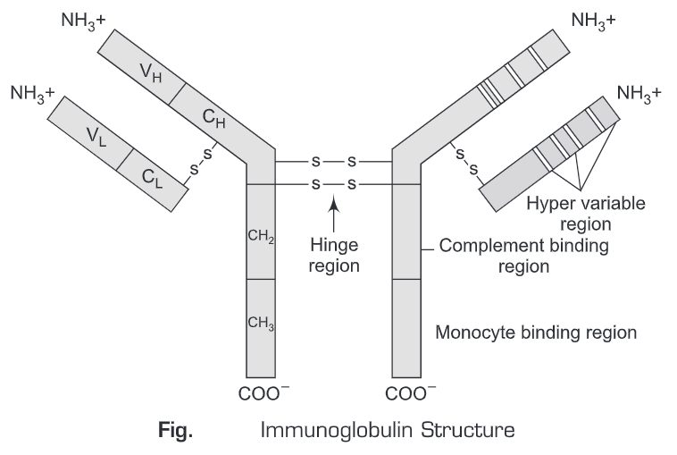
- Heavy and Light Chains: All immunoglobulins contain four chain structure as their basic unit. They contain two identical light chains (23kD) and two identical heavy chains (50-70kD)
- Disulfide Bonds: There are two types of disulfide linkages in immunoglobulins:
- Inter-chain disulfide bonds: The heavy and light chains and the two heavy chains are held together by inter-chain disulfide bonds and by non-covalent interactions The number of inter-chain disulfide bonds varies among different immunoglobulin molecules.
- Intra-chain disulfide bonds: Each polypeptide chain also contains intra-chain disulfide bonds.
- Variable (V) and Constant (C) Regions: When the amino acid sequences of many different heavy chains and light chains were compared, it became clear that both the heavy and light chain could be divided into two regions based on variability in the amino acid sequences.
These are the:
(i) Light Chain – VL (110 amino acids) and CL (110 amino acids)
(ii) Heavy Chain – VH (110 amino acids) and CH (330-440 amino acids)
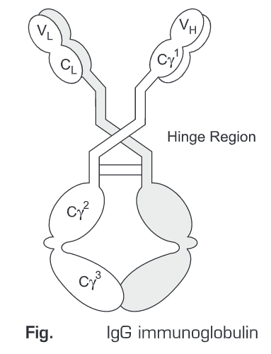
- Hinge Region: This is the region in which the arms of the antibody molecule form a Y structure. There is some flexibility in the molecule at this point therefore, it is known as hinge region.
- Domains: Domains are generally predicted by three-dimensional images of the immunoglobulin molecule. Studies show that it is not a straight chain rather it is folded into globular regions each of which contains an intra-chain disulfide bond. These regions are called domains.
(i) Light Chain Domains – VL and CL
(ii) Heavy Chain Domains – VH, CH1- CH3 (or CH4)
- Oligosaccharides: Carbohydrates are attached to the CH2, domain in most immunoglobulins. However, in some cases carbohydrates may also be attached at other locations.

Structure of the Variable Region
- Hypervariable (HVR) or complementarity determining regions (CDR)-Hypervariable regions or the complementarity determining regions are defined as the regions in which comparisons of the amino acid sequences of the variable regions of antibody show that most of the variability resides in these regions. Antibodies with different specificities (i.e. different combining sites) have different complementarity determining regions while antibodies of the exact same specificity have identical complementarity determining regions (i.e. CDR is the antibody combining site). Complementarity determining regions are found in both the H and the L chains.
- Framework regions-The regions which are present between the complementarity determining regions in the variable region are called the framework regions. Based on similarities and differences in the framework regions the immunoglobulin heavy and light chain variable regions can be divided into groups and subgroups. These represent the products of different variable region genes.
Immunoglobulin Fragments: Structure/Function Relationships
Determining the structure/function of the immunoglobulin fragments produced by proteolytic digestion has proven very useful.
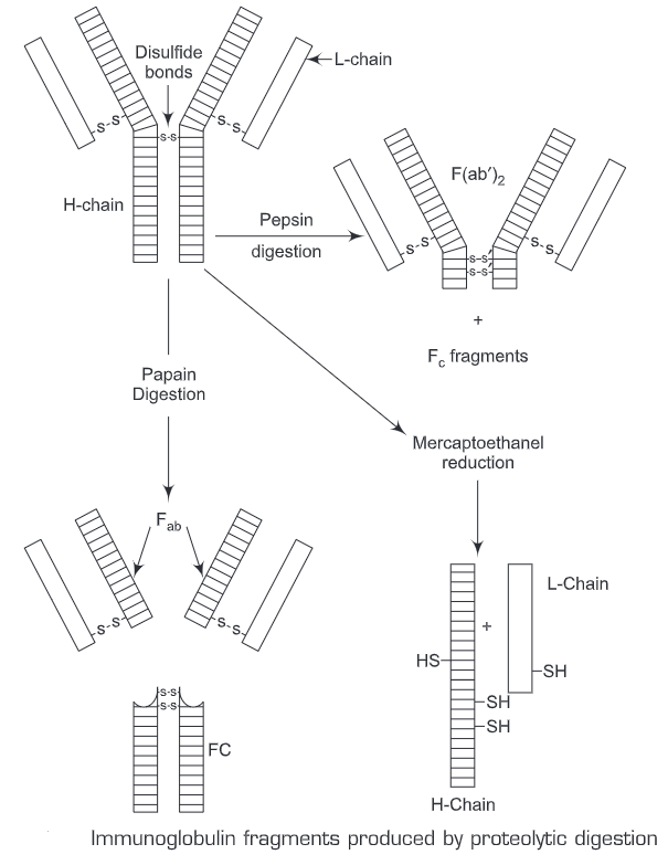
- Fab –When the immunoglobulin molecule is digested with papain, it breaks the hinge region before the H-H inter-chain disulfide bond which results in the formation of two identical fragments that contain the light chain and the VL, and CH2 domains of the heavy chain.
Antigen binding – These fragments were called the Fab fragments because they contained the antigen-binding sites of the antibody. Each Fab fragment is monovalent whereas the original molecule is divalent. The combining site of the antibody is created by both VH and VL. An antibody is able to bind a particular antigenic determinant because it has a particular combination of VH and VL. Different combinations of a Vj, and V, result in antibodies that can bind a different antigenic determinant.
- Fe –When the immunoglobulin is digested with papain, it produces a fragment that contains the remainder of the two heavy chains each containing a CH2 and CH3domain. This fragment was called Fc because it can be easily crystallized.
Effector functions – The effector functions of immunoglobulins are mediated by this part of the molecule. Different functions are mediated by the different domains in this fragment.
- F (ab’)2-Treatment of immunoglobulins with pepsin results in cleavage of the heavy chain after the H-H inter-chain disulfide bonds resulting in a fragment that contains both antigen binding sites. This fragment was called F(ab’)2because it is divalent. The Fc region of the molecule is digested into small peptides by pepsin. The F(ab’)2 binds antigen but it does not mediate the effector functions of antibodies.
Types of Human Immunoglobulin
- Immunoglobulin Classes:Immunoglobulins are divided into five different classes which are based on differences in the amino acid sequences in the constant region of the heavy chains. All immunoglobulins have similar heavy chain constant regions within the same regions. The main difference can be detected by sequence studies or more commonly by serological means.
- IgG – Gamma heavy chains
- IgM – Mu heavy chains
- IgA – Alpha heavy chains
- IgD – Delta heavy chains
- IgE – Epsilon heavy chains
- Immunoglobulin Subclasses:The immunoglobulins are further divided into subclasses based on small differences in the amino acid sequences in the constant region of the heavy chains. All immunoglobulins within a subclass will have very similar heavy chain constant region amino acid sequences. Again these differences are most commonly detected by serological means.
- IgG Subclasses
(a) IgG1 – Gamma 1 heavy chains
(b) IgG2 – Gamma 2 heavy chains
(c) IgG3 – Gamma 3 heavy chains
(d) IgG4 – Gamma 4 heavy chains
- IgA Subclasses
(a) IgA1 – Alpha 1 heavy chains
(b) IgA2 – Alpha 2 heavy chains
- Immunoglobulin Types:Another way of classifying immunoglobulins is through the type of light chain which they contain. Light chains are based on differences in the amino acid sequence in the constant region of the light chain. These differences are detected by serological means.
- Kappa light chains
- Lambda light chains
- Immunoglobulin Subtypes:The light chains can also be divided into subtypes based on differences in the amino acid sequences in the constant region of the light chain.
- Lambda subtypes
(a) Lambda 1
(b) Lambda 2
(c) Lambda 3
(2) Lambda 4
- Heterogeneity –Immunoglobulins considered as a population of molecules are normally very heterogeneous because they are composed of different classes and subclasses each of which has different types and subtypes of light chains. In addition, different immunoglobulin molecules can have different antigen-binding properties because of different VH; and VLregions.
Different Classes of Immunoglobulin
Billions of different variable regions are made and the general structure of antibodies falls into just five classes and this is based upon the type of heavy chain present in the antibody. In mammals, there are five classes of antibodies, IgA, IgD, IgE, IgG, and IgM, each with its own class of heavy chain. In addition, there are a number of subclasses of IgG and IgA immunoglobulins; for example, there are four human IgG subclasses (IgGl, IgG2, IgG3, and IgG). Each class (and subclass) has characteristic properties of its own. Except for their variable regions, all the immunoglobulins within a class have about 90% homology in their amino acid sequences, but only 60% homology exists between classes (eg., IgG and IgA).
- IgG – IgGis the most abundant immunoglobulin present in serum constituting about 80% of the total serum Ig. There are four human IgG subclasses that are distinguished by differences in Y-chain sequence and numbered according to their decreasing average serum concentrations – IgGl, 1gG2, 1gG3, and IgG4. The lgG isotype (except for subclass IgG2) is the only class of immunoglobulin that can pass through the placenta and enabling the mother to transfer her immunity to the fetus. Except for the IgG3 subclass, with a half-life of 7 days, the half-life of IgG is approximately 23 days, which is the longest half-life of all immunoglobulin isotypes.
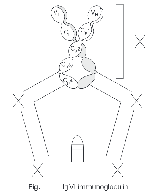
- IgM –IgM is the first class of antibody made by a developing B cell. It accounts for 5%-10% of the total serum Ig. IgM is also found on the surface of mature B cells together with IgD Monomeric IgM, has a molecular weight of 180,000, and is expressed as a membrane-bound antibody on B cells. IgM is secreted by plasma cells as a pentameric molecule, composed of five such units, each of which unit consists of two light and two heavy chains, which all are joined together by additional disulfide bonds between the Fc portions and by a polypeptide chain termed as the J chain. The J chain is synthesized in the B cell or plasma cell. The half-life of the IgM molecule is approximately 5 days. IgM is the first immunoglobulin class produced in a primary response to an antigen, and it is also the first Ig to be synthesized by the neonate. IgM is more efficient than IgG in activating complement. Complement activation requires two Fc regions in close proximity, and the pentameric structure of a single molecule of IgM fulfills this requirement.
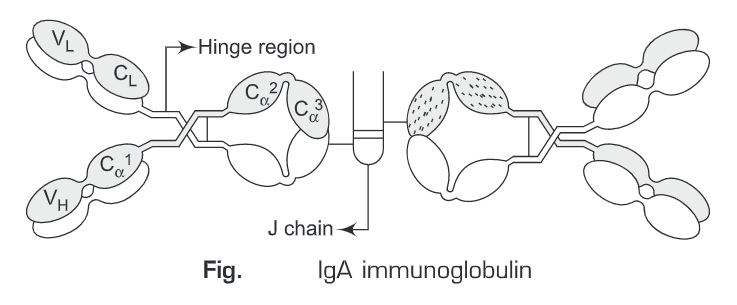
- IgA- 2IgA constitutes 10-15% of the total Ig in serum. It is the predominant Ig class in external secretions such as breast milk, saliva, tears, and mucus of the bronchial, genitourinary and digestive tracts. The IgA class of immunoglobulins contains two subclasses: IgA1 (93%) and IgA2 (7%). Serum IgA has a half-life of 5.5 days. The IgA present in serum is predominantly monomeric. The IgA of external secretions, called secretory IgA, consists of a dimer or tetramer with a J-chain polypeptide and a polypeptide chain called the secretory component. The daily production of secretory IgA is the maximum among other Ig. It is the major immunoglobulin found in the colostrum of milk in nursing mothers, and it may provide the neonate with a major source of protection against pathogens during the first few weeks after birth.
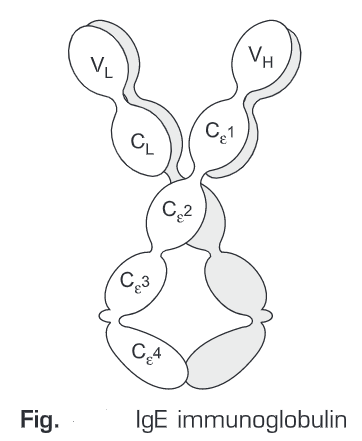
- IgE- IgE mediates the immediate hypersensitivity reactions that are responsible for the symptoms of hay fever, asthma, hives, and anaphylactic shock. IgE binds to Fe receptors on the membranes of blood basophils and tissue mast cells. Cross-linkage of receptor-bound IgE molecules by antigen (allergen) induces degranulation of basophils and mast cells; as a result a variety of pharmacologically active mediators giving rise to allergic manifestations. IgE-mediated degranulation is necessary for anti-parasitic defense.
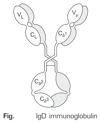
- IgD –IgD constitutes 0.2% of the total Ig in serum. IgD together with IgM, is the major membrane-bound Ig expressed by mature B-cells. It is thought to function in the activation of B cells by Ag. No biological effector function has been identified for IgD.
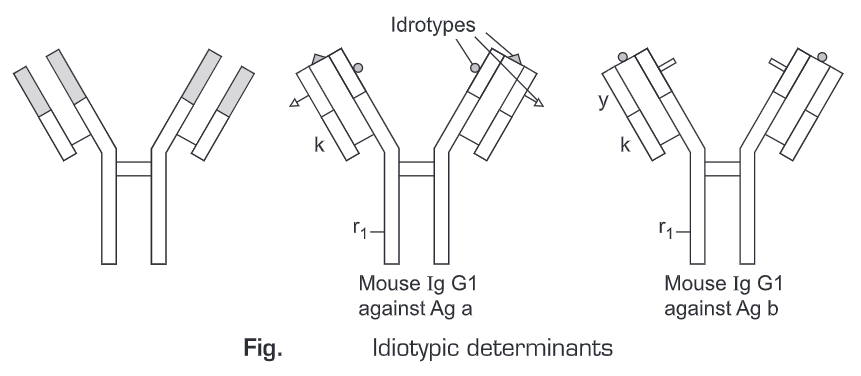
Antigenic Determinants on Immunoglobulins
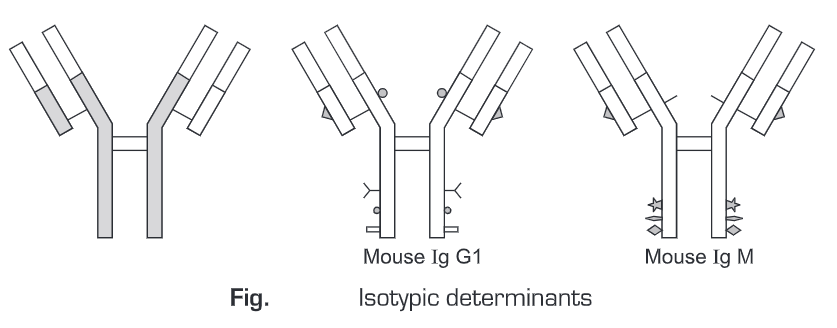
Since antibodies are glycoproteins, they can themselves function as potent immunogens to induce an antibody response. The antigenic determinants, or epitopes on Ig molecules fall into three major categories which are located in characteristic portions of the molecule.
- Isotype-An antibody class is determined by the constant region sequence of the heavy chain. The five human isotypes, designated IgA, IgD, IgE, IgG, and IgM, exhibit structural and functional differences. Isotypic determinants are constant-region determinants that collectively define each heavy-chain class and sub-class within a species. Each isotype is encoded by a separate constant region gene, and all members of a species carry the same constant-region genes. Different species inherit different constant-region genes and therefore express different isotypes. Therefore, when an antibody from one species is injected into another species, the isotype determinants will be recognized as foreign, inducing an antibody response to the isotypic determinants on the foreign antibody.
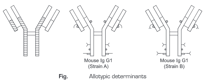
- Allotype –It is based on genetic differences among individuals. It depends on the existence of allelic forms of the same gene. These alleles encode minor amino-acid differences, called allotypic determinants that occur in some but not all, members of a species. As a result of allotypy, a heavy or light chain constituent of any immunoglobulin can be present in some members of a species and absent in others. This situation contrasts with that of immunoglobulin classes or subclasses, which are present in all members of a species.
- Idiotype –The unique amino-acid sequence of the variable domains of a given antibody can function not only as an antigen-binding site but also as a set of antigenic determinants. The idiotypic determinants are generated by the conformation of the heavy- and light-chain variable regions. Each individual antigenic determinant of the variable region is referred to as an idiotype.