Meninges and cerebrospinal fluid
Meninges
The brain and spinal cord are surrounded by three connective tissue membranes, the meninges. The subarachnoid space, filled with cerebrospinal fluid, separates the pia mater and arachnoid mater. Superficial cerebral blood vessels, invested by arachnoid mater, run through the subarachnoid space and their branches penetrate the brain becoming surrounded by a cuff of pia mater that extends as far as the capillaries. The perivascular (Virchow–Robin) space between the vessel wall and the pia mater is continuous with the subarachnoid space. Here the passive exchange of water and solutes across the pia mater keeps the CSF in equilibrium with brain extracellular fluid. At the cerebral capillaries the pia mater is lost and the single layer of capillary endothelial cells, with their basement membrane, are covered by glial cells. Expanded regions of the subarachnoid space are cisterns.
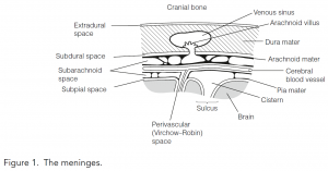
The dura mater is a thick tough outer layer with venous sinuses running through it. Small herniations of the arachnoid mater called arachnoid villi protrude through the dura into the venous sinuses. Here bulk flow of CSF into the blood occurs via mesothelial tubes in the arachnoid villi that act as valves, closing when the pressure in the venous sinus exceeds that of subarachnoid space to prevent reflux of blood into the CSF.
The subdural space is a potential space between the dura mater and the arachnoid mater. It is traversed by cerebral veins entering the venous sinuses in the dura.
Cerebrospinal fluid circulation
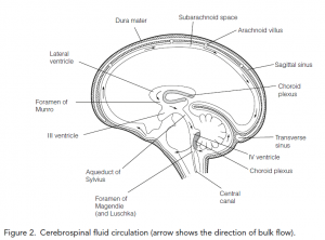
Cerebrospinal fluid (CSF) is actively secreted by choroid plexuses situated in the lateral, third, and fourth ventricles. The flow of CSF is from the lateral ventricles through the foramen of Munro into the third ventricle, and then through the aqueduct of Sylvius into the fourth ventricle. From here it drains via three orifices into the subarachnoid space. Here it equilibrates with extracellular fluid in the perivascular spaces. Finally, it is dumped into the venous sinuses via the arachnoid villi.
CSF secretion
The choroid plexus consists of a cuboidal epithelium covering a core of a highly vascular pia mater. In adult humans, CSF is secreted at about 500 cm3 day-1 into a steady-state volume of 100–150 cm3. Of this, about 30 cm3 is in the ventricles and the rest is in the subarachnoid space. Cerebrospinal fluid is turned over about every 5–7 hours.
The choroid plexus secretes some substances and absorbs others, most by active transport mechanisms. In this way, it acts as a selective interface between blood and CSF, the blood–CSF barrier. The result is that by comparison with blood plasma CSF has somewhat higher Na+, Cl-, and HCO3- concentrations but lower K+, urea, glucose, and amino acid concentrations. Although the protein concentration of CSF is about 1000-fold lower than blood plasma, its higher ionic concentration gives the two fluids the same osmolality.
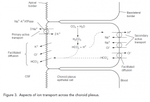
Aspects of ion transport across the blood–CSF barrier is shown in Figure 3. Na+, K+-ATPase on the apical border of the epithelial cell pumps sodium into the CSF. This generates a sodium gradient that drives two secondary active transport mechanisms bringing Na+ across the basolateral border; Na+–H+ exchange and a Na+–Cl- symport. The Cl- influx in turn drives a Cl-–HCO3 – antiport. Bicarbonate brought into the cell in this way is added to that formed intracellularly by hydration of CO2, a reaction greatly accelerated by the high levels of carbonic anhydrase present in the choroid plexus. The bicarbonate diffuses via an apical anion transporter into the CSF.
CSF and meningeal functions
The functions of the CSF are metabolic and mechanical. By equilibrating with brain extracellular fluid unwanted metabolites (choline, dopamine, and serotonin metabolites, urea, creatinine, and K+) are transported into the blood, either via arachnoid villi or choroid plexuses. There are three mechanical effects which are protective:
- Because the subarachnoid space is a fluid-filled compartment in which the brain floats, the effective weight of the brain is reduced from about 1350 g to about 50 g.
- Adjustments to CSF and meninges prevent changes in intracranial pressure due to alterations in cerebral blood flow. When blood flow increases, CSF is squeezed from the ventricles into the subarachnoid space around the spinal cord. Here the dura mater is more elastic and stretches to accommodate the rise in volume. Longer-term increases in intracranial pressure can be offset by a rise in CSF flow into the venous sinuses through the arachnoid villi.
- The meninges (particularly the dura) support the brain and the CSF reduces the force with which the brain impacts the inside of the cranium when the head moves.