What is a Neuron?
- Definition: A neuron is a specialized cell in the nervous system that receives, processes, and transmits electrical and chemical signals to communicate with other cells.
- Role in the Nervous System: Neurons are the primary cells responsible for relaying information, coordinating bodily functions, and enabling cognitive processes.
- Types of Neurons: Based on function, neurons are classified into:
- Sensory Neurons: Transmit sensory information from receptors to the central nervous system (CNS).
- Motor Neurons: Carry signals from the CNS to muscles or glands to initiate action.
- Interneurons: Facilitate communication between neurons within the CNS.
- Significance: Neurons enable rapid communication, forming complex networks that underpin all nervous system activities, from reflexes to higher cognitive functions like memory and learning.
Structure of a Neuron
The structure of a neuron is uniquely adapted to its role in signal transmission. Neurons have a distinct morphology with specialized components that allow them to receive, process, and transmit signals.
Key Structural Features
- Polarized Structure: Neurons have a receiving end (dendrites) and a transmitting end (axon), creating a directional flow of information.
- Membrane: The neuron’s plasma membrane is selectively permeable, maintaining an electric potential critical for signal transmission.
- Specialized Organelles: Neurons contain organelles like mitochondria and endoplasmic reticulum, supporting high metabolic demands.
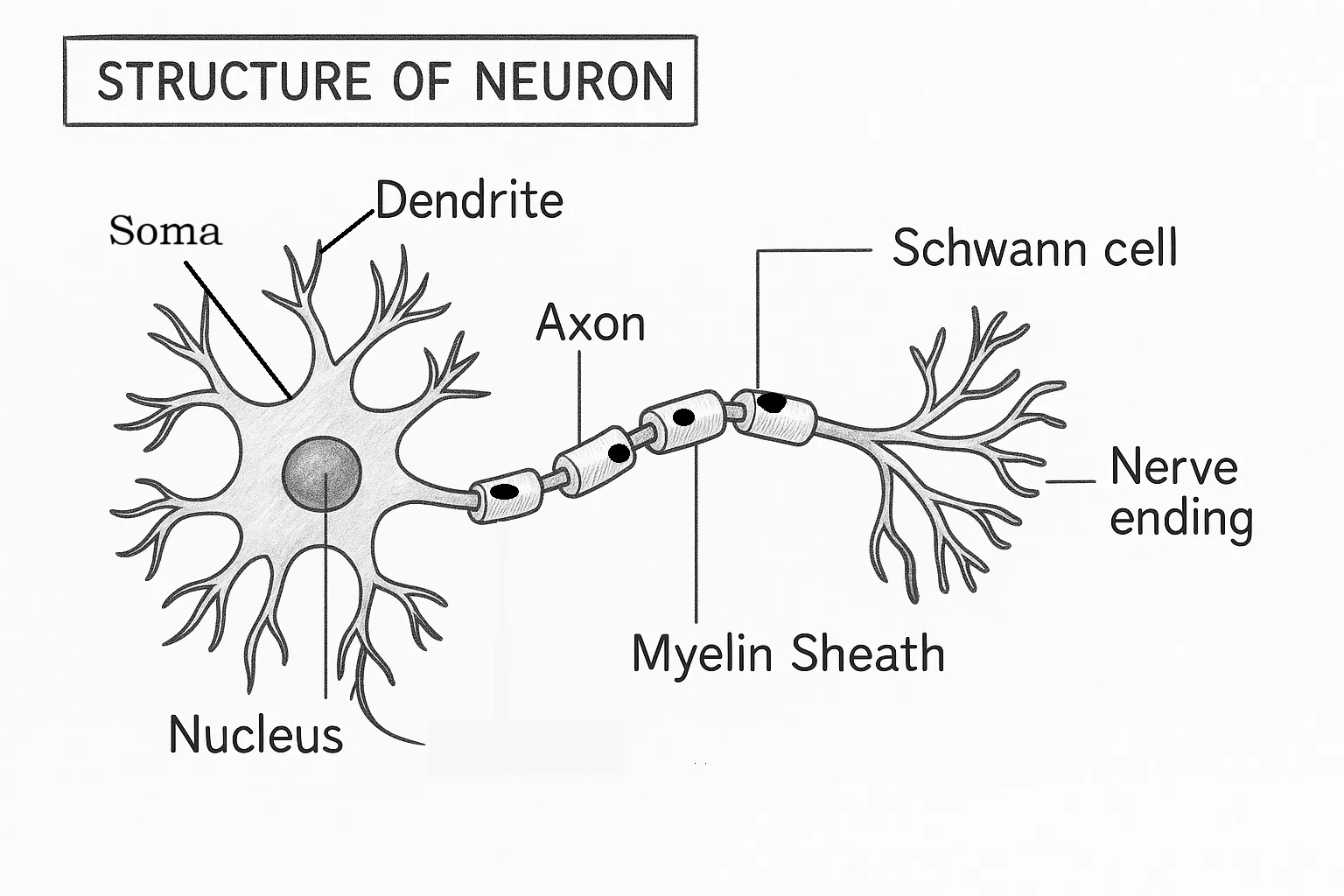
Main Parts of a Neuron
Neurons consist of three primary regions: the cell body, dendrites, and axon, each with specific roles.
Table 1: Main Parts of a Neuron
| Part | Description | Key Features |
|---|---|---|
| Cell Body (Soma) | The central part containing the nucleus and organelles. | Houses genetic material and metabolic machinery; integrates incoming signals. |
| Dendrites | Branched extensions that receive signals from other neurons or sensory receptors. | Highly branched; contain receptors for neurotransmitters; short in length. |
| Axon | A long, slender projection that conducts electrical impulses away from the soma. | May be myelinated for faster signal conduction; terminates in axon terminals. |
Detailed Parts of a Neuron
Let’s break down the components of a neuron in greater detail, exploring their substructures and specific roles.
1. Cell Body (Soma)
- Definition: The soma is the neuron’s metabolic center, containing the nucleus and essential organelles.
- Components:
- Nucleus: Stores DNA and regulates gene expression for protein synthesis.
- Mitochondria: Provide energy (ATP) for neuronal activity.
- Endoplasmic Reticulum (ER):
- Rough ER: Synthesizes proteins, such as ion channels and receptors.
- Smooth ER: Regulates calcium levels and lipid synthesis.
- Golgi Apparatus: Modifies and packages proteins for transport.
- Nissl Bodies: Clusters of rough ER and ribosomes, prominent in neurons, for protein synthesis.
- Function:
- Maintains neuronal health and functionality.
- Integrates incoming signals from dendrites to determine whether to generate an action potential.
2. Dendrites
- Definition: Dendrites are tree-like extensions that receive input from other neurons or sensory receptors.
- Structure:
- Highly branched to maximize surface area for synaptic connections.
- Contain dendritic spines, small protrusions that increase connectivity.
- Equipped with receptors for neurotransmitters.
- Types:
- Apical Dendrites: Found in pyramidal neurons, extending from the apex of the cell body.
- Basal Dendrites: Extend from the base of the soma, common in cortical neurons.
- Function:
- Receive and integrate chemical signals (neurotransmitters) from other neurons.
- Convert chemical signals into electrical signals (graded potentials).
3. Axon
- Definition: The axon is a long, cable-like structure that transmits electrical impulses (action potentials) away from the soma.
- Structure:
- Axon Hillock: The region where the axon originates from the soma; critical for initiating action potentials.
- Axon Shaft: The elongated portion, which may be myelinated or unmyelinated.
- Myelin Sheath: A fatty insulating layer formed by glial cells (oligodendrocytes in CNS, Schwann cells in PNS), speeding up signal transmission.
- Nodes of Ranvier: Gaps in the myelin sheath where action potentials are regenerated.
- Axon Terminals: The distal ends of the axon, forming synapses with target cells.
- Function:
- Conducts action potentials over long distances.
- Releases neurotransmitters at synapses to communicate with other cells.
4. Synapses
- Definition: Synapses are specialized junctions where neurons communicate with other neurons, muscles, or glands.
- Components:
- Presynaptic Terminal: The axon terminal of the transmitting neuron, containing neurotransmitter-filled vesicles.
- Synaptic Cleft: A narrow gap between the presynaptic and postsynaptic cells.
- Postsynaptic Membrane: The receiving cell’s membrane, containing receptors for neurotransmitters.
- Types:
- Chemical Synapses: Use neurotransmitters to transmit signals (most common).
- Electrical Synapses: Allow direct passage of ions via gap junctions (faster but less common).
- Function:
- Facilitate communication between neurons or between neurons and effector cells.
- Enable signal amplification, modulation, or inhibition.
Table 2: Subcomponents of a Neuron
| Subcomponent | Location | Function |
|---|---|---|
| Nucleus | Cell Body | Stores genetic material; regulates protein synthesis. |
| Dendritic Spines | Dendrites | Increase surface area for synaptic connections; modulate signal strength. |
| Axon Hillock | Base of Axon | Initiates action potentials based on integrated signals. |
| Myelin Sheath | Axon | Insulates axon to speed up action potential conduction. |
| Nodes of Ranvier | Axon | Sites of action potential regeneration for efficient signal transmission. |
| Synaptic Vesicles | Axon Terminals | Store and release neurotransmitters into the synaptic cleft. |
Functions of Neurons
Neurons perform a variety of functions critical to the nervous system’s operation. These functions are enabled by their ability to generate, propagate, and transmit electrical and chemical signals.
1. Signal Reception
- Role: Neurons receive signals from sensory receptors or other neurons via dendrites.
- Mechanism:
- Dendrites detect neurotransmitters released by presynaptic neurons.
- Binding of neurotransmitters to receptors generates graded potentials (small changes in membrane potential).
- Significance: Allows neurons to gather sensory or synaptic input, forming the basis for information processing.
2. Signal Integration
- Role: The soma and axon hillock integrate incoming signals to determine whether to fire an action potential.
- Mechanism:
- Graded potentials from dendrites summate (add up) in the soma.
- If the combined signal exceeds a threshold at the axon hillock, an action potential is triggered.
- Types of Summation:
- Temporal Summation: Multiple signals from the same source arriving in quick succession.
- Spatial Summation: Signals from multiple sources arriving simultaneously.
- Significance: Enables decision-making at the cellular level, filtering out weak or irrelevant signals.
3. Signal Conduction
- Role: Neurons transmit electrical impulses (action potentials) along the axon to communicate with distant cells.
- Mechanism:
- Action potentials are generated at the axon hillock when the membrane potential depolarizes to a threshold.
- The impulse travels along the axon via changes in ion concentrations (sodium and potassium ions).
- In myelinated axons, action potentials “jump” between Nodes of Ranvier (saltatory conduction), increasing speed.
- Significance: Ensures rapid and reliable transmission of signals over long distances.
4. Signal Transmission
- Role: Neurons transmit signals to other neurons, muscles, or glands via synapses.
- Mechanism:
- Arrival of an action potential at the axon terminal triggers the release of neurotransmitters.
- Neurotransmitters cross the synaptic cleft and bind to receptors on the postsynaptic cell, generating a new signal.
- Significance: Enables communication between cells, coordinating complex physiological and behavioral responses.
5. Plasticity and Learning
- Role: Neurons adapt their structure and function in response to experience, supporting learning and memory.
- Mechanism:
- Synaptic Plasticity: Changes in synapse strength, such as long-term potentiation (LTP) or depression (LTD).
- Structural Plasticity: Growth of new dendritic spines or axonal branches in response to activity.
- Significance: Underpins cognitive functions like learning, memory, and adaptation to new environments.
Table 3: Functions of Neurons
| Function | Description | Key Mechanism |
|---|---|---|
| Signal Reception | Receiving sensory or synaptic input via dendrites. | Neurotransmitter binding to dendritic receptors generates graded potentials. |
| Signal Integration | Summating inputs to decide whether to fire an action potential. | Temporal and spatial summation at the soma and axon hillock. |
| Signal Conduction | Transmitting electrical impulses along the axon. | Action potentials propagate via ion channel activity; enhanced by myelination. |
| Signal Transmission | Relaying signals to other cells via synapses. | Neurotransmitter release at axon terminals. |
| Plasticity | Adapting structure/function for learning and memory. | Synaptic and structural changes in response to activity. |
How Neurons Work Together
Neurons rarely function in isolation; they form complex networks to process and transmit information. Here’s how they collaborate:
- Neural Circuits: Groups of interconnected neurons that process specific types of information (e.g., visual or motor circuits).
- Reflex Arcs: Simple circuits involving sensory neurons, interneurons, and motor neurons for rapid responses (e.g., knee-jerk reflex).
- Neural Networks: Large-scale networks in the brain that integrate sensory, cognitive, and motor functions.
- Neurotransmitter Systems: Different neurons release specific neurotransmitters (e.g., dopamine, serotonin), influencing mood, movement, and cognition.
Clinical Relevance of Neurons
Understanding neurons is crucial for addressing neurological disorders:
- Neurodegenerative Diseases:
- Alzheimer’s Disease: Loss of neurons and synapses in the cortex and hippocampus impairs memory.
- Parkinson’s Disease: Degeneration of dopamine-producing neurons affects movement.
- Neuropsychiatric Disorders:
- Depression: Altered neurotransmitter signaling (e.g., serotonin) affects mood regulation.
- Schizophrenia: Dysregulated neural circuits in the prefrontal cortex and limbic system.
- Injury and Repair:
- Stroke: Loss of blood supply damages neurons, impairing function.
- Neuroplasticity: The brain’s ability to reorganize neural connections aids recovery after injury.
FAQs About Neurons
1. What is the difference between a neuron and a nerve?
- A neuron is a single nerve cell, while a nerve is a bundle of axons from multiple neurons, often encased in connective tissue, that transmits signals to specific body regions.
2. How many neurons are in the human brain?
- The human brain contains approximately 86 billion neurons, though estimates vary slightly.
3. Can neurons regenerate?
- In the adult human CNS, neuron regeneration is limited, but some peripheral neurons can regenerate axons. Neurogenesis (new neuron formation) occurs in specific brain regions, like the hippocampus, but is minimal in adults.
4. What is the role of myelin in neurons?
- Myelin insulates axons, speeding up action potential conduction via saltatory conduction and protecting axons from damage.
5. How do neurons communicate with each other?
- Neurons communicate primarily via chemical synapses, where neurotransmitters released by one neuron bind to receptors on another, triggering a response.
References
- Kandel, E. R., Schwartz, J. H., & Jessell, T. M. (2021). Principles of Neural Science. McGraw-Hill Education. Link
- Purves, D., Augustine, G. J., Fitzpatrick, D., et al. (2018). Neuroscience. Sinauer Associates. Link
- National Institute of Neurological Disorders and Stroke. (2023). Brain Basics: The Life and Death of a Neuron. Link
- Bear, M. F., Connors, B. W., & Paradiso, M. A. (2020). Neuroscience: Exploring the Brain. Wolters Kluwer. Link
- Lodish, H., Berk, A., Zipursky, S. L., et al. (2021). Molecular Cell Biology. W.H. Freeman. Link