Human Brain
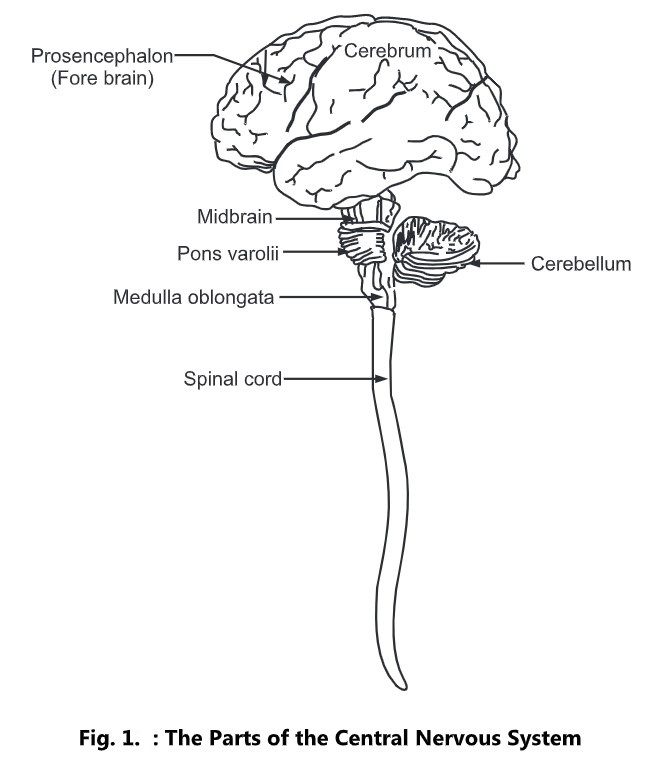
The brain lies within the cranial cavity. It constitutes about 1/5th of the body weight. It is composed of the following parts:
- Cerebrum or forebrain
- Midbrain
- Pons Varolii
- Medulla oblongata
- Cerebellum or hindbrain
Cerebrum
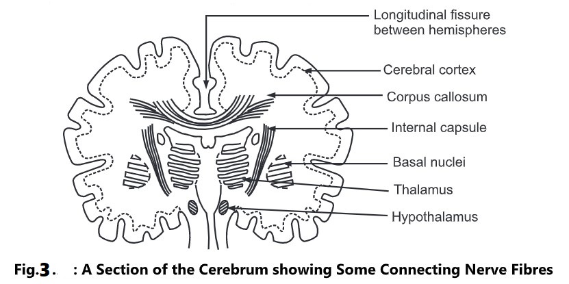
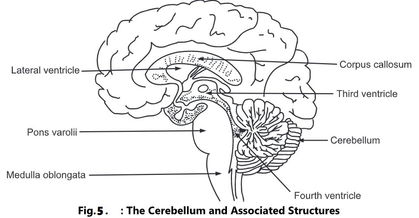
It is the largest part of the brain and occupies anterior and middle part of the cranial cavity. It is divided by a deep cleft into right and left cerebral hemispheres. Deep within the brain, the hemispheres are connected by a mass of nerve fibres called corpus callosum. The superficial part of the cerebrum is composed of nerve cell body, forming cerebral cortex.
The cerebral cortex shows many enfolding of varying depth. The exposed areas of the fold are called as gyro or convolutions and are separated by sulci or fissure. The convolutions help in increasing surface area of the cerebrum.
Each hemisphere of the cerebrum is divided into the following lobes:
- Frontal
- Parietal
- Temporal
- Occipital
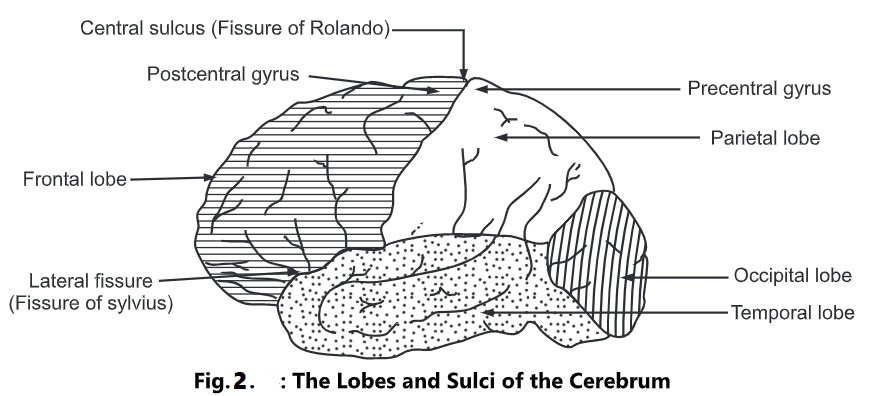
The boundaries of the lobes are marked by deep sulci; called as central, lateral and parietal occipital sulci.
Within the cerebrum the lobes are connected by masses of nerve fibres, or tracks, which constitute white matter of the brain. Various afferent (incoming) and efferent (outgoing) fibres linking the different parts of the brain and spinal cord are as follows:
- Arcuate (association) fibres: These fibres connect different parts of a cerebral hemisphere by extending from one gyro to another.
- Commissural fibres These fibres connect corresponding areas of the two cerebral hemispheres. The largest commissural is called as Corpus callosum.
- Projection fibres: These fibres connect the cerebral cortex with grey matter of lower parts of the brain and with the spinal cord. Internal capsule is one of the projection fibres. It lies deep within the brain between the basal ganglia and the thalamus. All nerve impulses passing to and from the cerebral cortex are carried by fibres that form the internal capsule. Motor fibres within the internal capsule form the pyramidal tracts (corticospinal tracts) that decussate at the medulla oblongata.
Functions of the Cerebrum
- Mental activities involved in memory, intelligence, sense of responsibility; thinking, reasoning, moral sense, and learning.
- Sensory perception, including the perception of pain, temperature, touch; sight, hearing, taste and smell.
- Initiation and control of voluntary muscle contraction.
Functional Areas of the Cerebrum
Functional areas of the cerebrum are divided into sensory and motor area. Anatomically there is a mixture of both these functions.
Motor Areas
The precentral area lies in the frontal lobe immediately anterior to the central sulcus. The nerve cells are pyramid-shaped and they initiate contraction of voluntary muscles. A nerve fibre from a pyramid-shaped cell passes downwards through the internal capsule to the medulla oblongata where it crosses to the opposite side and descends in the spinal cord.
As a result a motor area of the right hemisphere of the cerebrum controls voluntary muscle movements on the left side of the body and vice-versa. The neuron with its cell body in the cerebrum is termed as the upper motor neuron. The neuron with its cell body in the spinal cord is termed as lower motor neuron. Damage to either of neurons may result in paralysis.
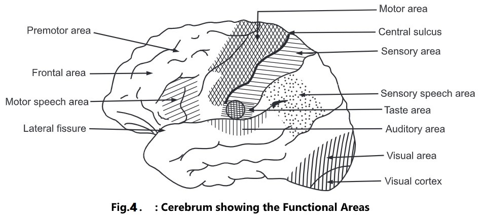
The premotor area lies in the frontal lobe immediately anterior to the motor area. The cells are supposed to exert controlling influence over the motor area, ensuring orderly movements. In the lower part of this area just about the lateral sulcus, there is a group of nerve cells known as the motor speech area which controls movements necessary for speech. It is dominant in the left hemisphere in right-handed people and vice-versa.
The frontal area extends anteriorly from the premotor area to include the remainder of frontal lobe. It is a large area. It is highly developed in human beings than in other animals. It is responsible for the behaviour, character and emotional states of the individual.
Sensory Areas
The post central (Sensory) area lies behind the central sulcus. In this area, sensations of pain, temperature, pressure; touch, knowledge of muscular movement, and the position of joints are perceived. The sensory area of the right hemisphere receives impulses from the left side of the body and vice-versa.
The parietal area lies behind the post-central area and includes the greater part of the parietal lobe of the cerebrum. Its functions are supposed to be associated with obtaining and retaining accurate knowledge of objects. The sensory speech area is situated in the lower part of the parietal lobe and extends into the temporal lobe. The spoken word is perceived here. The auditory (hearing) area lies immediately below the lateral sulcus within the temporal lobe. The cells receive and interpret impulses transmitted from the inner ear by the vestibulocochlear (auditory) nerves. The olfactory (smell) area lies deep within the temporal lobe where impulses from the nose via the olfactory nerves are received and interpreted. The taste area lies just above the lateral sulcus in the deep layers of the sensory area. In this area, impulses from special nerve endings in taste buds in the tongue and in the lining of the cheeks, palate, and pharynx are perceived as taste. The visual area lies behind the parietal-occipital sulcus and includes the greater part of the occipital lobe. The optic nerves (nerves of the sense of sight) pass from the eye to this area. It receives the impulses as visual impressions.
Other Areas
Deep within the cerebral hemispheres, there are groups of cell bodies termed as nuclei. They act as relay stations where impulses are passed from one neuron to the next in a chain.
Following are important masses of grey matter.
- Basal nuclei
- Thalamus
- Hypothalamus
- Basal nuclei: This area lies deep within the cerebral hemispheres. It is supposed to influence skeletal muscle tone. If control is inadequate or absent, movements are jerky, clumsy and uncoordinated.
- Thalamus: It consists of two masses of nerve cells and fibres situated within the cerebral hemispheres just below the corpus callosum, one on each side of the third ventricle. Sensory input from the skin, viscera, and special sense organs is transmitted to the thalamus before redistribution to the cerebrum.
- Hypothalamus: It is composed of a number of groups of nerve cells. It is situated below and in front of the thalamus, immediately above the pituitary gland. It is linked to the posterior lobe of the pituitary gland by nerve fibres and to the anterior lobe by a complex system of blood vesicles. It controls the output of hormones from both lobes of the gland. It controls the following additional functions.
- Autonomic nervous system.
- Hunger and thirst.
- Body temperature.
- Emotional reactions: pleasure, fear, etc.
- Sexual behaviour.
- Biological clocks or circadian rhythms, e.g. sleeping and waking cycles.
Brain Stem
Mid brain: It is situated around the cerebral aqueduct between the cerebrum above and the Pons varolii below. It consists of groups of nerve cells and nerve fibres. It connects the cerebrum with lower parts of the brain and the spinal cord. The nerve cells act as relay stations for the ascending and descending nerve fibres.
Pons varolii: It is situated in front of the cerebellum, below the midbrain and above the medulla oblongata. It consists of nerve fibres forming a bridge between the two hemispheres of the cerebellum, and of fibres passing between the higher levels of the brain and the spinal cord. Few groups of cells within the Pons act as relay stations. Some of them are associated with the cranial nerves. The nerve cells in the Pons lie deep and the nerve fibres are on the surface.
Medulla oblongata: It extends from the Pons varolii above and is continuous with the spinal cord below. It is shaped like a pyramid with its base upwards and it lies within the cranium above the foramen magnum. Its anterior and posterior surfaces are marked by central fissures. Some cells of medulla oblongata constitute relay stations for sensory nerves passing from the spinal cord to the cerebrum. Following vital centres which are associated with autonomic reflex activity lie in its deeper structure.
- Cardiac centre
- Respiratory centre
- Vasomotor centre
- Reflex centres of vomiting, coughing, sneezing and swallowing.
Medulla oblongata has the following special features:
-Decussating of the pyramids: Motor nerves descending from the motor area in the cerebrum to the spinal cord in the pyramidal or corticospinal tracts, cross from left side to right and vice versa. These tracts are the main pathways for impulses to voluntary, i.e. skeletal muscles.
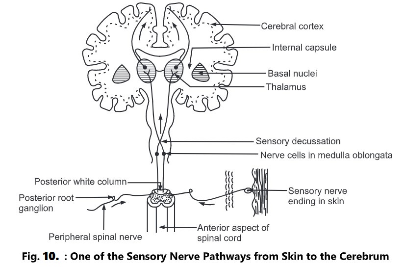
– Sensory decussating: Some of the sensory nerves ascending to the cerebrum from the spinal cord cross from left side to right and vice-versa in the medulla oblongata. Few other sensory nerves decussate at the level of spinal cord.
– The cardiac centre: It controls the rate and force of cardiac contraction. Sympathetic and parasympathetic nerve fibres starting from the medulla pass to the heart.
– The respiratory centre: It controls the rate and depth of respiration. From this center, nerve impulses pass to the Phrenic and intercostal nerves which stimulate contraction of the diaphragm and intercostal muscles, thus initiating inspiration. The center is stimulated by excess CO2 or by deficiency of O2 in blood supply. Variation in concentrations of CO2,/O2 is conveyed by nerve impulses from the chemoreceptors in the carotid bodies.
– The vasomotor centre: It controls the diameter of the blood vessels. It has special control over small arteries and arterioles which influence a large proportion of smooth muscle fibres in their walls, eg. the sources of stimulation of the vasomotor center, the arterial baroreceptors, body temperature and emotions such as sexual excitement and anger. Pain usually causes vasoconstriction; however, severe pain may cause vasodilatation, a fall in blood pressure and
fainting.
– Reflex centres: When irritating substances are present in the stomach or respiratory tract nerve impulses pass to the medulla oblongata, stimulating the reflex centers; this in turn initiates reflex actions of vomiting, coughing and sneezing.
Reticular Formation
It is collection of neurons in the core of the brainstem, surrounded by neural pathways which pass nerve impulses between the brain and the spinal cord. It constantly receives information from the brain and the spinal cord and transmits it in descending and ascending tracts. It has the following functions:
- Co-ordination of skeletal muscle activity associated with voluntary motor movement and the maintenance of balance.
- Co-ordination of activity control by the autonomic nervous system, e.g. cardiovascular, respiratory, and gastrointestinal activity.
- Selective awareness functions through the Reticular Activating System (RAS). This can selectively block or pass sensory information to the cerebral cortex.
Cerebellum
It is situated behind the Pons varolii and immediately below the posterior portion of the cerebrum occupying the posterior cranial fosse.
It has two hemispheres, separated by a narrow median strip called as vermis. On the surface of the cerebellum lies grey matter, while the white matter lies deep inside. The cerebellum has got the following functions:
- It is concerned with the co-ordination of voluntary muscular movement, posture, and balance.
- It controls and co-ordinates the movements of various groups of muscles ensuring smooth, even, precise actions.
- It co-ordinates activities associated with the maintenance of the balance and equilibrium of the body.
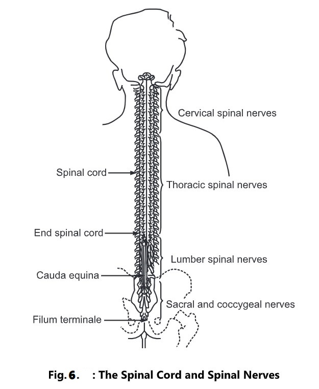
Spinal Cord
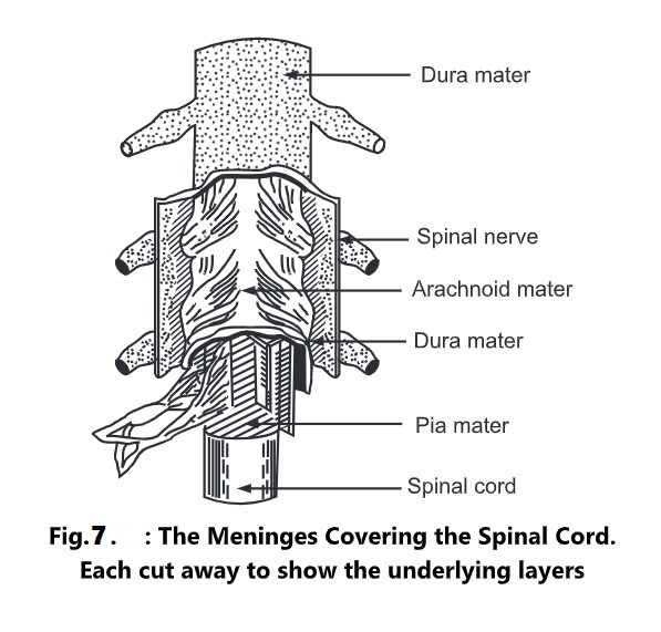
It is the elongated and almost cylindrical part extending from brain just below medulla oblongata. It is suspended in the vertebral canal and is surrounded by meninges and cerebrospinal fluid.
It extends from the first cervical vertebra to the lower border of first lumbar vertebra. It is approximately 45 cm long and about the thickness of the little finger. Except for the cranial nerves, the spinal cord is the nervous tissue link between the brain and the rest of the body. Motor nerves originating from the brain descends through the spinal cord and supply to various organs and tissues at appropriate levels of the cord. Sensory nerves from different organs and tissues enter and pass upwards to the brain via the spinal cord. Some activities like spinal reflexes are independent of the brain. In such cases motor action is decided and implemented at the level of spinal cord itself. In order to facilitate spinal reflexes, there are extensive neuron connections between sensory and motor neurons at the same or different levels in the spinal cord.
A cross-section of the spinal cord shows that it is composed of grey matter in the center and is surrounded by white matter supported by neuralgia.
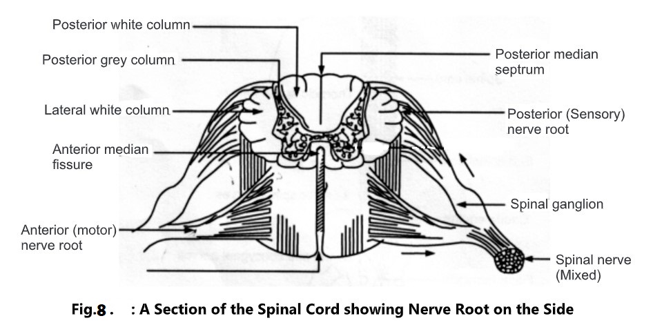
The arrangement of grey matter in the spinal cord resembles the shape of the letter H, having two anterior, two posterior and two lateral columns. The area of grey matter lying transversely is the transverse commission and is pierced by the central canal. The canal extends from the fourth ventricle in the brain and contains cerebrospinal fluid. The spinal cord consists of the following cell bodies:
- Sensory cells receiving impulses from the periphery of the body.
- Lower motor neurons transmitting impulses to the skeletal muscles.
- Connector neurons, linking sensory and motor neurons at the same or different levels.
Posterior columns of grey matter are composed of cell bodies which are stimulated by sensory impulses from the body. The nerve fibres of these cells form white matter and transmit the sensory impulses to the brain.
Anterior columns of grey matter are composed of the cell bodies of the lower motor neurons; they are stimulated by the axons of the upper motor neurons or by the cell bodies of connector neurons. All sensory nerve fibres pass through posterior root ganglia. These ganglia promote onward movement of nerve impulses from the periphery.
The white matter of the spinal cord is arranged in three columns or tracts; anterior, posterior and lateral. These tracts are formed by sensory nerve fibres ascending to the brain, motor nerve fibres descending from the brain and fibres of connector neurons.
Sensory Nerve Tracts in the Spinal Cord
Following are two main sources of sensation transmitted to the brain via the spinal cord:
1. The Skin: Sensory nerve endings in the skin are called as cutaneous receptors. They are stimulated by pain, heat, cold; touch and pressure. The sensory impulses are passed to the opposite hemisphere of the cerebrum and the sensation is perceived in that region.
2. The tendons, muscles, and joints: Sensory nerve endings in these structures are called proprioceptors and are stimulated by stretch. Along with impulses from the eyes and ears, they are associated with maintenance of balance and posture and with perception of the position of the body in space.
Motor Nerve Tracts in the Spinal Cord
Neurons which transmit nerve impulses away from the brain are termed as motor neurons. Stimulation of these neurons results in the following actions:
- Contraction of skeletal muscles.
- Contraction of smooth muscles and the secretion by glands controlled by nerves of the autonomic nervous system.
Movements of Voluntary Muscles:
The contraction of the muscles moving the joints is a voluntary act. Stimulus for this act originates at the level of consciousness in the cerebrum. Some impulses are originated in the midbrain, brainstem and cerebellum. This activity occurs below the level of consciousness and is associated with coordination of muscle activity.
Efferent nerve impulses are transmitted from the brain via bundles of nerve fibres for tracts through the spinal cord. There are two types of tracts in the spinal cord:
- Pyramidal (corticospinal)
- Extra pyramidal
The motor fibres forming pyramidal tracts travel through the internal capsule and are the main pathway for impulses to skeletal muscles. Those motor fibres which do not pass through the internal capsule form the extra pyramidal tracts and have connections with many parts of the brain including the basal nuclei and the thalamus.
The upper motor neuron has its cell (Betz’s cell) in the precentral sulcus of the cerebrum. The axons pass through the internal capsule, Pons and medulla. In this spinal cord they form the lateral corticospinal tracts and these fibres terminate in close association with the cells of lower motor neurons in the anterior columns of grey matter. The axons of these upper motor neurons make up the pyramidal tracts and decussate in the medulla oblongata, forming the pyramids.
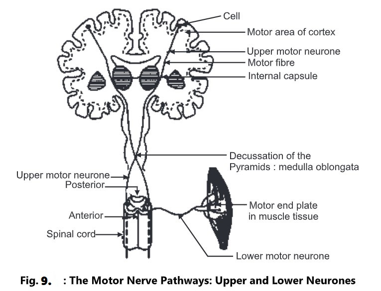
The lower motor neuron has its cell in the anterior horn of grey matter in the spinal cord. Its axon emerges from the spinal cord by the anterior root, joins with the incoming sensory fibres and forms the mixed spinal nerve. At its termination in muscle the axon branches into a variable number of tiny fibres which form motor end-plates. Each motor end-plate has close association with a sensitive area on the wall of a muscle fibre. The motor end-plates of each nerve fibre and the muscle fibre form a motor unit.
At the end of motor unit, acetylcholine is secreted after stimulation of the nerve. Acetylcholine is termed as a neurotransmitter. The secretion of this neurotransmitter is responsible for the contraction of smooth or skeletal muscles.
The lower motor neuron is the final common pathway for the transmission of nerve impulses to skeletal muscles. The cell of this neuron is influenced by various upper motor neurons. Some of these neurons stimulate the cells while others inhibit them. A balance between these two actions results in smooth, co-ordinated muscle movement. A part of this movement may be voluntary and a part of it may be involuntary in nature.
Movements of Involuntary Muscles
Upper motor neurons have their cells in the brain at level below the cerebrum that is in the midbrain, brainstem, and cerebellum or spinal cord. They influence muscle activity in relation to the maintenance of posture and balance, the co-ordination of muscle movement, and the control of muscle tone.
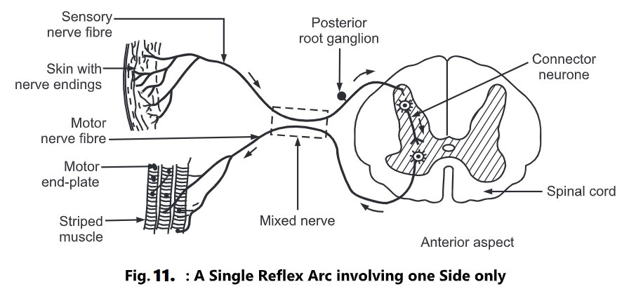
Spinal reflexes consist of three elements:
- Sensory neurons.
- Connector neurons.
- Lower motor neurons.
In the reflex arc, only one of the elements exists. A reflex action is an immediate motor response to a sensory stimulus. Many connector and motor neurons may be stimulated by efferent impulses from a small area of skin. These, in turn, stimulate many connector and lower motor neurons in the cord which results in the contraction of many skeletal muscles of different organs. Reflex action takes place very quickly. It is of a protective type; they may be inhibited occasionally.
In stretch reflexes, only two neurons are involved. The cell of the lower motor neuron is stimulated by the sensory neuron. There is no connector neuron involved in it, e.g. knee jerk.
By tapping the tendon just below the knee when it is bent, the sensory nerve endings in the tendon, and in the thigh muscles are stretched. This initiates a nerve impulse which passes into the spinal cord to the cell of the lower motor neuron in the anterior column of grey matter on the same side. As a result, the thigh muscles suddenly contract, and the foot kicks forward.