Tissues of the Human Body
Introduction
A tissue is a group of cells having a common embryonic origin that functions together to carry out specialized activities. Tissues may be hard (bone), semisolid (fat), or liquid (blood) in their consistency. Tissues may vary with respect to the types of cells present, their arrangement, and the type of fibres present.
Histology:
It is the branch of science that deals with the study of tissues.
Types of Body Tissues:
According to the structure and functions, tissues can be divided as follows:
(1) Epithelial tissue: It covers the body surfaces, lines hollow organs, body cavities, and ducts.
(2) Connective tissue: It protects and supports the body and its organs. It acts as an energy store (reserves fat) and provides immunity.
(3) Muscle tissue: It generates the physical force needed to generate body heat.
(4) Nervous tissue: It detects the changes inside and outside the body environment and generates nerve impulses responsible for muscular contractions and glandular secretions.
1. Epithelial Tissue: Types, Structure, and Function
- An epithelial tissue or epithelium consists of cells arranged in continuous sheets, in either single or multiple layers.
- It covers body surfaces and lines hollow organs, body cavities, and ducts. It also forms glands.
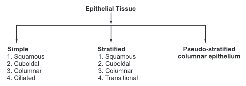
Types of Epithelial Tissue
Functions of Epithelial Tissue:
- It protects the underlying tissue from friction and injury.
- It secret certain chemical substances that are utilized by the body.
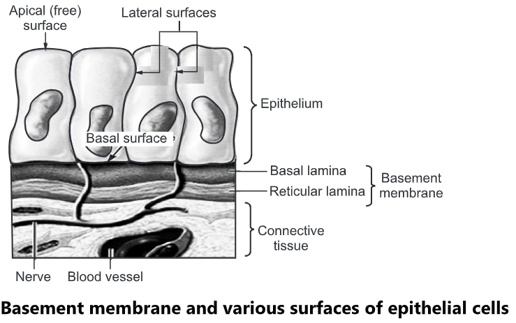
- On the basal side, the epithelial cells are supported by a basement membrane called as basal lamina.
- Below the basal lamina lies the capillary bed which provides the epithelium with the required nutrients and eliminates the waste products.
Surfaces of Epithelial Cells:
- Apical (free) surface: It faces the body surface. It may contain a body cavity, the lumen of an internal organ that receives cell secretions. It may contain cilia or microvilli.
- Lateral surface: It faces the adjacent cells on either side. It may contain tight junctions, adherens junctions, desmosomes, and/or gap junctions.
- Basal surface: It is present opposite to the apical surface. In multiple layers of epithelial cells, the apical layer is the most superficial layer of cells, and the basal layer is the deepest layer of cells.
Basement Membrane:
- It is a thin extracellular layer that consists of two layers, the basal lamina, and the reticular lamina.
- The basement membrane is a point of attachment and support for the overlying epithelial tissue.
- Basal lamina: It is closer to and secreted by the epithelial cells. It contains proteins such as laminin and collagen as well as glycoproteins and proteoglycans.
- Reticular lamina: It is closer to the underlying connective tissue and contains proteins such as collagen produced by connective tissue.
Simple Epithelium
It is made up of a single layer of cells and is divided into four types:
(a) Simple squamous epithelium
(b) Simple cuboidal epithelium
(c) Simple columnar epithelium
(d) Simple ciliated epithelium
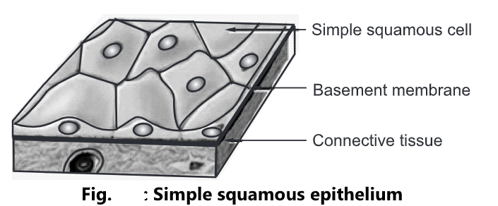
Simple Squamous Epithelium:
- It consists of a single layer of flat cells arranged on the basement membrane. The nucleus of each cell is oval or spherical and is centrally located.
- Location: It lines the heart, blood vessels, lymphatic vessels, air sacs of lungs and
the glomerular capsule of kidneys. - Functions: It performs the function of filtration (such as blood filtration in the kidneys), diffusion (such as diffusion of oxygen onto blood vessels of the lungs), and osmosis
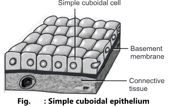
Simple Cuboidal Epithelium:
- It is made up of a single layer of cube-shaped cells arranged on the basement membrane.
- The nucleus of each cell is spherical in shape and is centrally located.
- Location: It lines the kidney tubules, pancreas and also forms the covering of ovaries.
- Functions: It performs the function of protection to the underlying tissues, secretion, and absorption of filtered substances.
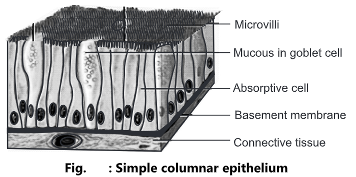
Simple Columnar Epithelium:
- It is made up of a single layer of rectangular cells arranged on the basement membrane.
- The nucleus of each cell is oval in shape and is located near the base of the cell. Mucous secreting columnar epithelium cells are called goblet cells.
- Location: It is the most abundant cell in the body. They are found in the nasal passage, eye, digestive system, reproductive system, ears, and buccal cavity.
- Functions: It performs the function of protection, secretion, providing sensory input, absorption, and transporting nutrients in the small intestine.
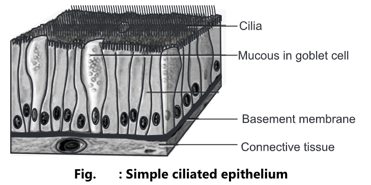
Simple Ciliated Epithelium:
- It is made up of columnar epithelial cells with many hair-like projections at the top called cilia. The nucleus of each cell is oval in shape and is located near the base of the cell. The cells are connected by tight junctions and desmosomes. The mucous secreting columnar epithelium cells are called as goblet cells.
- Location: It lines the upper respiratory tracts, uterine (fallopian) tubes, uterus, and central canal of the spinal cord.
- Functions: The cilia move the mucous and other substances by ciliary action. This prevents the adherence of any particulate matter such as bacteria, thus preventing infection. In the uterine tubes, the cilia propel the ova towards the uterus, and in the respiratory passages, they propel mucous towards the throat.
Stratified Epithelium
- It is made up of multiple layers of cells.
- It is further divided into four types.
- Stratified squamous epithelium
- Stratified cuboidal epithelium
- Stratified columnar epithelium
- Transitional epithelium
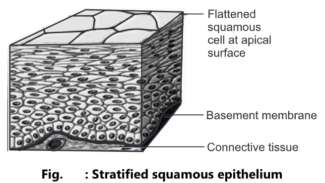
Stratified Squamous Epithelium:
- It is composed of more than one layer of cells having different shapes.
- The cells in the apical layer are flat and those present in deep layers vary in shape from cuboidal to columnar.
- As the cell grows their blood supply is restricted and they become dehydrated, shrunken, and harder.
- These issues exist in two forms keratinized and non-keratinized.
- In keratinized stratified squamous epithelium, the apical layer and several deep layers are dehydrated and contain a layer of keratin protein, a tough, fibrous protein that helps to protect the skin and underlying tissues from heat, microbes, and chemicals.
- In non-keratinized stratified squamous epithelium, keratin is absent from the apical layer and they remain moist.
- Location: Keratinized cells from the superficial layer of skin, Non-keratinized cells line the wet surfaces such as the lining of mouth, esophagus, part of epiglottis, vagina, and also covers the tongue.
- Functions: It gives protection against mechanical friction and chemical damage.
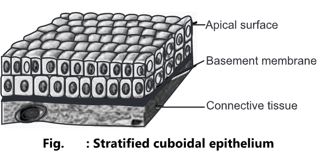
Stratified Cuboidal Epithelium:
- It is made up of two or more layers of cells. The cells in the apical layer are cuboidal in shape.
- Location: It lines the ducts of the sweat gland, male urethra, uterus, and anus.
- Functions: It plays an important role in protection, secretion, and absorption.
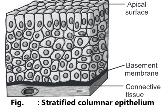
Stratified Columnar Epithelium:
- It is made up of several layers of irregularly shaped cells. In the apical layer, columnar cells are present.
- Location: It lines part of the urethra, large excretory ducts of glands, and conjunctiva of the eye.
- Functions: It performs the function of protection and secretion.
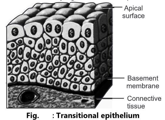
Transitional Epithelium:
- It consists of many layers of pear-shaped cells. The cells are variable in appearance.
- In a relaxed state, it looks like stratified cuboidal epithelium, and when stretched the cells become squamous shaped.
- Location: It lines the hollow organs such as the uterus and urinary bladder.
- Functions: It protects the underlying structure and permits the distension of organs.
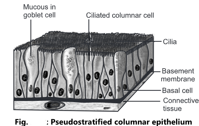
Pseudostratified Columnar Epithelium
- The cells are columnar in shape having a hair-like structure at the top called cilia.
- The nucleus is oval in shape and is present at different positions.
- All the cells are attached to the basement membrane, but not all reach the apical surface.
- Location: It lines the airways of the upper respiratory tract; pseudostratified non-ciliated columnar epithelium lines larger ducts of many glands, epididymis, and male urethra.
- Function: It functions in the secretion and movement of mucous by ciliary action.
Glandular Epithelium
- A gland may consist of a single cell or a group of cells.
- They are specialized cells that secrete substances into ducts.
- The glands are classified into endocrine or exocrine glands on the basis of their secretions.
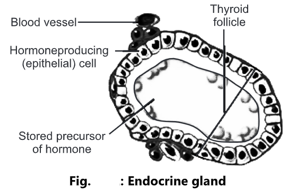
Endocrine gland:
- They are ductless glands.
- The secretions of endocrine glands enter the interstitial fluid and then diffuse directly into the bloodstream.
- These secretions are called hormones that regulate metabolic and physiological activities of the body in order to maintain homeostasis.
- Location: The pituitary gland, pineal gland, thyroid gland, parathyroid gland, adrenal glands, pancreas, ovaries, testes, and thymus are examples of endocrine glands.
- Function: The function of the endocrine gland is the production of hormones that regulate various metabolic and physiological activities.
- The pituitary gland secretes the human growth hormone responsible for the normal growth of individuals.
- The pineal gland secretes the melatonin hormone responsible for maintaining circadian rhythms and seasonal functions.
- The thyroid gland secretes T3 and T4 which are responsible for maintaining the normal functioning of the thyroid gland.
- The pancreas secretes the insulin hormone responsible for controlling the blood sugar level.
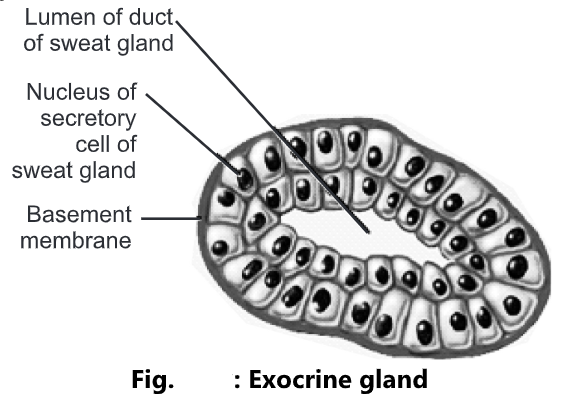
Exocrine Gland:
- These are the glands that possess ducts.
- The secretions of these glands are released into ducts that empty onto the skin surface or the lumen of a hollow organ.
- The secretions of exocrine glands include mucous, sweat, oil, earwax, saliva, and digestive enzymes.
- The secretory products of exocrine glands are released into the ducts.
- Examples of exocrine glands include sudoriferous (sweat) glands, which produce sweat that helps to reduce the body temperature, and salivary glands which secrete saliva.
- Location: They are present in the sweat gland, sebaceous gland, earwax glands, salivary glands, and pancreas
- Function: The function of exocrine glands in the production of sweat, oil, earwax, saliva, or digestive enzyme.
- Exocrine glands are classified as unicellular or multicellular on the basis of a number of cells.
Unicellular glands:
- These are composed of a single epithelial cell.
- They lack ducts.
- They secret their products directly on the surface of body cavities. For example, Goblet cells secrete mucous directly onto the apical surface of the intestinal tract.
Multicellular glands:
- These are composed of more than one cell.
- The epithelium grows down from the surface into the underlying tissues to form a simple or compound tube.
- It consists of an epithelium-derived duct and a secretory unit that is surrounded by connective tissue. E.g. Sudoriferous, sebaceous (oil), and salivary glands.
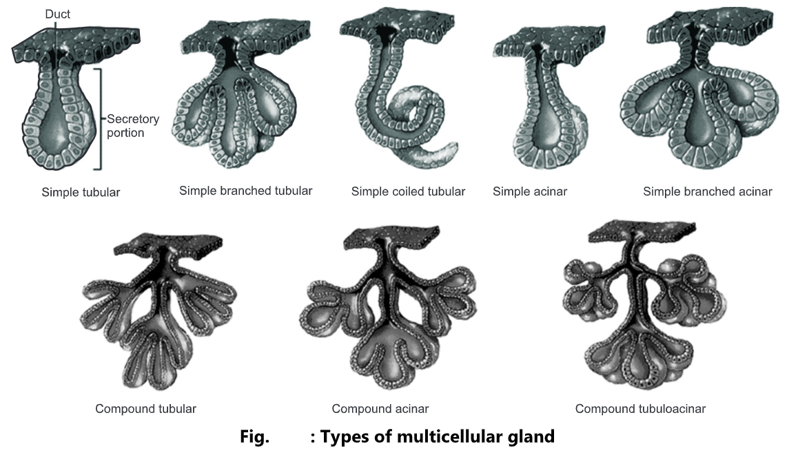
Types of Multicellular Gland:
On the basis of the structure of their ducts, multicellular exocrine glands are classified as:
Simple gland: If the duct of the gland does not branch it is called a simple gland.
Compound gland: If the duct of the gland is branched it is called a compound gland.
Tubular gland: If the duct of the gland is tubular in shape it is called a tubular gland.
Acinar gland: If the duct of the gland is rounded it is called as acinar gland.
Tubuloacinar gland: If the duct of the gland is both tubular and rounded it is called a tubuloacinar gland.
On the basis of their secretory unit, multicellular exocrine glands are classified as;
Simple glands:
- Simple tubular: Tubular secretory part is straight and attaches to a single unbranched duct. Example: Glands in the large intestine.
- Simple branched tubular: Tubular secretory part is branched and attaches to a single unbranched duct. Example: Gastric glands.
- Simple coiled tubular: Tubular secretory part is coiled and attaches to a single unbranched duct. Example: sweat glands.
- Simple acinar: Secretory portion is rounded and attaches to a single unbranched duct. Example: Glands of the urethra.
- Simple branched acinar: Rounded secretory part is branched and attaches to a single unbranched duct. Example: Sebaceous glands.
Compound Glands:
- Compound tubular: Secretory portion is tubular and attaches to a branched duct. Example: Bulbo-urethral glands.
- Compound acinar: Secretory portion is rounded and attaches to a branched duct. Example: Mammary glands.
- Compound tubuloacinar: Secretory portion is both tubular and rounded and attaches to a branched duct. Example: Acinar glands of the pancreas.
2. Connective Tissue: Types, Structure and Function
- It is the most abundant and widely distributed tissue system in the body.
- It binds together, supports and strengthens other body tissues as well as protects and insulates internal organs.
- It is made up of fibres, cells and ground substances.
Fibres:
Three types of fibres are embedded in the extracellular matrix between the cells.
These fibres strengthen and support connective tissues.
(a) Collagen fibres:
These are very strong and allow tissue flexibility. These are made up of protein collagen. It is the most abundant protein making up about 25% and 35% of the total body protein. Collagen fibre is often presented in parallel bundles. It is found in bone, cartilage, tendons and ligaments.
(b) Elastic fibres:
These fibres are smaller in diameter. These are made up of protein elastin surrounded by a glycoprotein named fibrillin which gives strength and stability to tissue. Elastic fibres have the ability to return to their original shape, a property called elasticity. These are found in the skin, lungs, arteries, veins, elastic cartilage, periodontal ligament and foetal tissue.
(c) Reticular fibres:
They consist of collagen protein arranged in fine bundles covered with glycoprotein. These are much thinner than collagen fibres. They give support and strength. These are found in the liver, bone marrow and lymphatic organs.
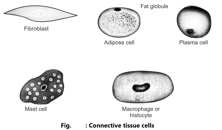
Cells:
Each cell consists of fibroblasts, macrophages, plasma cells, mast cells, adipocytes and white blood cells.
(a) Fibroblasts:
They are the chief cells of connective tissue. They are large, flat cells with branching processes.
(b) Macrophages:
These cells develop from monocytes, a type of white blood cells. There are two types of macrophages. These cells play an important role in the immune response. Fixed macrophages are present in particular tissue such as alveolar macrophages in lungs or spleen macrophages in spleen. Wandering macrophages have the ability to move throughout the tissue and gather at the site of infection to carry phagocytosis.
(c) Plasma cells:
A small cell that develops from a type of white blood cell is called β-lymphocytes. It takes an important part in the immune response. They are present in the gastrointestinal and respiratory tract, salivary glands, lymph nodes, spleen and red bone marrow.
(d) Mast cells:
They produce histamine that dilates the small blood vessels as a part of the inflammatory response.
(e) Adipocytes:
These are also called fat cells or adipose cells. They store fats. They are found deep in the skin and around the heart and kidneys.
(f) White blood cells:
In response to the inflammatory reaction, they migrate from the blood into connective tissue. E.g. Neutrophils gather at sites of infection and eosinophils migrate to the sites of allergic response.
Ground Substance:
- It is an amorphous gel-like substance present surrounding the cells.
- In the ground substance, cells and fibres are suspended.
- It supports the cells, binds them together, stores water and provides a medium through which substances are exchanged between blood and cells.
- It is primarily composed of water, glycosaminoglycan (hyaluronan), proteoglycans, glycoproteins, hyaluronic acid, chondroitin sulfate and dermatan sulfate.
Functions:
- It acts as an energy store.
- It provides protection to different body organs.
- It provides a structural framework to the body
- It connects different body tissues.
- It connects epithelial tissues to muscle fibres.
- It supplies hormones all over the body.
Classification of Connective Tissue:
Types of Connective tissues are as follows:
Loose Connective Tissue:
- Areolar connective tissue
- Adipose connective tissue
- Reticular connective tissue
Dense Connective Tissue:
- Dense regular connective tissue
- Dense irregular connective tissue
- Elastic connective tissue
Cartilage:
- Hyaline cartilage
- Fibrocartilage
- Elastic cartilage
Bone tissue:
Liquid Connective Tissue:
- Blood tissue
- Lymph
Loose Connective Tissue
- The fibres are loosely woven. It has a large proportion of ground substance. They are easily distorted. On distortion, they become tough and resist further deformation.
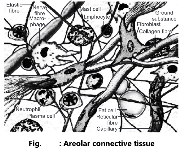
Areolar Connective Tissue:
- They form a loose network in the intercellular material and are not arranged in a particular pattern.
- It consists of collagen fibre, elastic fibres, reticular fibres and several kinds of cells such as fibroblasts, macrophages, plasma cells, adipocytes and mast cells embedded in ground substances.
- Location: It is present below the skin, fill the spaces between muscles, supports blood vessels and nerves in the alimentary canal.
- Yellow elastic fibres are found in arteries and white elastic fibres are found in kidneys and the brain.
- Functions: It gives strength, elasticity and support to tissue.
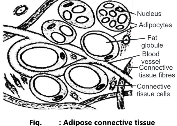
Adipose Connective Tissue:
- It consists of adipocytes that stores fats as a large centrally located droplet.
- Location: It is present in the subcutaneous layer deep in the skin, around the heart and kidneys and yellow bone marrow.
Functions:
- It prevents heat loss from the body.
- It acts as a reservoir of energy.
- It gives shape to the limbs and body
- It protects the underlying organ from injury.
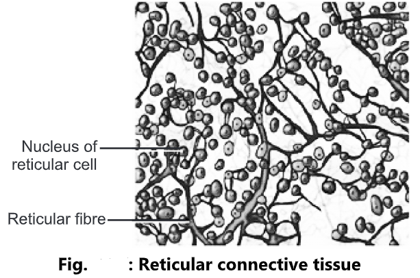
Reticular Connective Tissue:
- It consists of reticular fibres and reticular cells.
- Location: It is present in the supporting framework of the liver, spleen, lymph nodes, red bone marrow and is also found around blood vessels and muscles.
- Functions: It forms the stroma of organs, binds together smooth muscle tissue cells, filters and removes worn-out blood cells in the spleen and microbes in the lymph node.
Dense Connective Tissue
- In this, fibres are densely packed, the fibres content is higher and cell content is lower as compared to loose connective tissue.
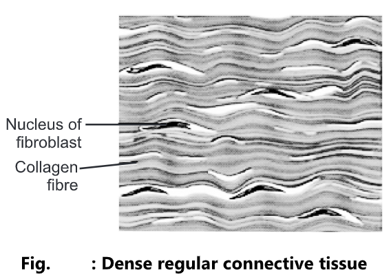
Dense Regular Connective Tissue:
- Bundles of collagen fibres are arranged in parallel patterns to provide strength to the tissue. Fibroblasts appear in rows between the fibres. It is silvery-white in colour and tough in nature.
- Location: It forms tendons (attach muscle to bone) and ligaments (attach bone to bone).
- Functions: It provides a strong attachment to structures.
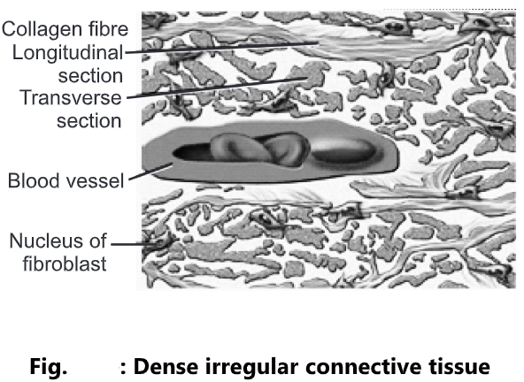
Dense Irregular Connective Tissue:
- It contains collagen fibres that are irregularly arranged and a few fibroblasts appear in rows between the fibres.
- Location: It is present in the tissue beneath the skin, the dermis of the skin, periosteum of bone, membrane capsules around kidneys, liver, testes, lymph node, pericardium of heart and heart valves.
- Functions: It provides strength to different organs.
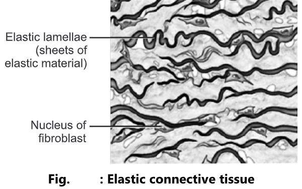
Elastic Connective Tissue:
- It consists of freely branching elastic fibres. Fibroblasts are present in spaces between fibres. It is yellowish in colour.
- Location: It is present in the lung tissues, wall of elastic arteries, trachea, bronchial tubes and vocal cords.
- Function: It allows the stretching of various organs.
Cartilage
It consists of a network of closely packed collagen fibres and elastic fibres embedded in a gelatinous substance called chondroitin sulfate of the ground substance. The cells of mature cartilage are called chondrocytes.
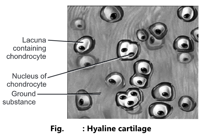
Hyaline Cartilage:
- It is bluish-white in colour. It consists of fine collagen fibres and many chondrocytes. It is the most abundant cartilage in the body.
- Location: It is present at the ends of long bones, anterior ends of ribs, nose, and part of larynx, trachea, bronchi, bronchial tubes, embryonic and fetal skeleton.
- Functions: It provides small surfaces for movement at joints, flexibility and support.
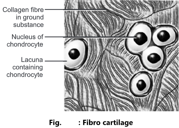
Fibro Cartilage:
- It is the strongest form of cartilage. The chondrocytes are scattered among the bundle of collagen fibres within the extracellular matrix.
- It is tough and slightly flexible.
- Location: It is present in the intervertebral disc.
- Functions: It covers and protects the bony structures of the body.
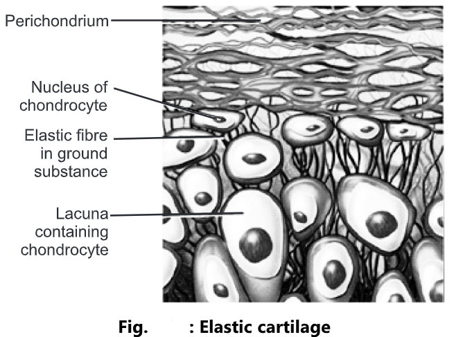
Elastic Cartilage:
- The chondrocytes are located within a threadlike network of elastic fibres within the extracellular matrix.
- Location: It is present in the pinna of the ear and top of the larynx.
- Functions: It provides strength and elasticity and maintains the shape of certain organs, such as the external ear.
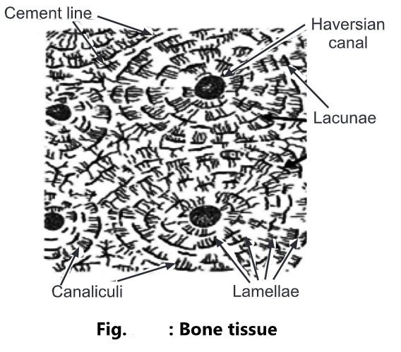
Bone
- It is the hardest connective tissue.
- It has a calcified matrix containing many collagen fibres.
- It is composed of 25% of water, 30% organic material and 45% inorganic salts.
- It is well vascularised.
- It is arranged in concentric ring structures called osteons.
- At the centre of the ring is a structure called as Haversian canal.
- Haversian canal system consists of:
- Central Haversian Channel: It contains blood vessels and nerves.
- Lamellae: Surrounding the central canal, concentric plates of bone are presently called lamellae.
- Lacunae: It contains mature bone cells called osteocytes.
- Canaliculi: Projecting from the lacunae are canaliculi, a network of minute canals containing the processes of osteocytes.
- Location: It is present in compact and spongy bone tissue.
Functions:
- To form a supporting framework for the body.
- To give protection to delicate organs.
- To form joints essential for locomotion of body.
- To form red blood cells in the red bone marrow.
- To provide a store of calcium salts.
- It gives support and maintains shape.
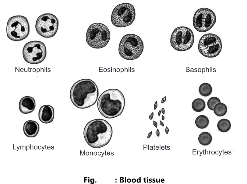
Blood
- It is a connective tissue with a liquid extracellular matrix called blood plasma.
- The blood cells are suspended in the blood plasma.
- It is composed of 55% plasma and 45% of cells.
- Blood plasma is a straw-coloured liquid in which the blood cells are suspended.
- Plasma is composed of 90-92% of water, 7% plasma proteins and clotting factors, and 1% of mineral salts, sugar, hormones and vitamins.
- Blood cells are of three types;
- Erythrocytes (RBC): These cells transport oxygen to body cells and remove carbon dioxide from them.
- Leucocytes (WBC): These cells are involved in phagocytosis, immunity and allergic reaction.
- Thrombocytes (Platelets): These cells participate in the process of blood clotting.
- Location: It is present within blood vessels (arteries, arterioles, capillaries, venules and veins) and within the chambers of the heart.
Functions:
- RBCs transport oxygen to body cells and remove carbon dioxide from them.
- WBCs are involved in phagocytosis, immunity and allergic reaction.
- Platelets participate in the blood clotting process.
3. Muscles Tissue: Types, Structure and Function
- Muscular tissue consists of elongated cells called muscle fibres that can use ATP to generate force.
- Three types of muscular tissues are present.
- Skeletal/striated/voluntary muscle tissue
- Cardiac muscle
- Smooth/non-striated/involuntary muscle tissue.
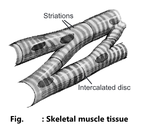
Skeletal Muscle Tissue
- The cells are cylindrical in shape.
- The fibres are parallel to each other.
- The length of muscle fibre is 30-40 cm.
- They have several nuclei located at the periphery.
- It shows alternate dark and light bands i.e. striations and hence the name is striated muscle.
- The muscles are attached to the bones hence called skeletal muscles.
- The activity of fibres is within one’s control hence, the name is a voluntary muscle.
- Location: It is usually attached to bone by tendons.
- Functions: It helps to give motion, posture, heat production and protection.
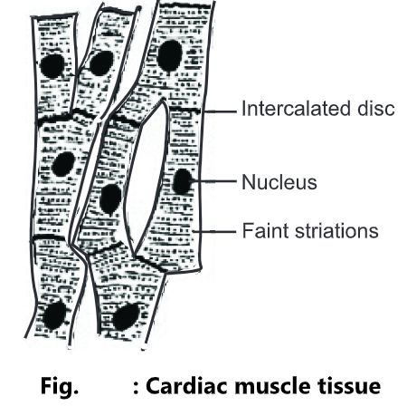
Cardiac Muscle Tissue
- It is present in the myocardium of the heart wall.
- It is striated but involuntary i.e. the activity of fibres is beyond one control hence called as involuntary muscle tissue.
- Each fibre is parallel to the other, branched and multinucleated.
- Two cardiac muscle fibres are attached by a thickened plasma membrane called an intercalated disc.
- The intercalated disc contains desmosomes as well as gap junctions.
- Location: It is present in the heart wall.
- Functions: It pumps blood to all parts of the body, contracts the atria and ventricles of the heart, causes rhythmic beating of the heart.
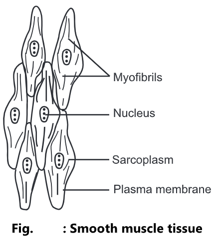
Smooth Muscle Tissue
- A smooth muscle fibre is usually small.
- It is thickest in the middle and tapering at the ends
- It contains a single, centrally located nucleus.
- The cells are spindle-shaped.
- Alternate light or dark bands are absent hence they are called smooth/non-striated.
- The activity of these fibres is beyond one’s control or wish and hence called involuntary.
- Location: It is present in the wall of blood vessels, wall of lymph vessels, alimentary tract, respiratory tract, urinary bladder and uterus.
- Functions: It gives motion (contraction of blood vessels, airways, propulsion of food through GIT, contraction of urinary bladder and gall bladder).
4. Nervous Tissue: Structure and Function
- It is made up of two types of nerves cells.
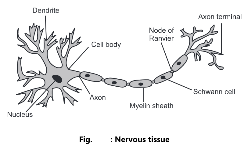
(a) Neurons:
- It is made up of a cell body, axons, dendrites and axon terminals.
- Cell body: It contains the nucleus and other organelles.
- Dendrites: These are input portions of neurons. These are usually short and highly branched forms tree-like structures. Each nerve cell contains many dendrites.
- Axon: Each nerve cell contains a single axon which is the thin, long and cylindrical process. It is the major output portion of a neuron that conducts the signal to effector organs. The axons are surrounded by a white, fatty substance called the myelin sheath. The unmyelinated regions between the myelin segments are called Nodes of Ranvier.
- Axon terminals: These are responsible for transmitting the signals.
- Location: It is present in the nervous system.
- Functions: It exhibits sensitivity to various types of stimuli, converts stimuli into nerve impulses (action potentials), and conducts nerve impulses to other neurons, muscle fibres, or glands.