Nose (Nasal Cavity)
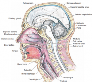
- The median nasal septum, consisting of bony and cartilaginous components, subdivides the nasal cavity into a right and a left nasal fossa, Each nasal fossa has anterior and posterior apertures, the naris and choana, respectively.
- In addition, each possesses four out pocketing termed the paranasal or accessory sinuses, a medial and a lateral wall, a floor, and a roof. The entrance into the nasal fossa immediately superior to the naris is a bilateral area of that cavity ringed by the greater alar cartilage termed the vestibule.
- The vestibule is lined by skin possessing vibrissae and sebaceous glands. The superior-most portion of the nasal fossa, specialized for olfaction, is the olfactory region, whereas the larger, inferior portion is the respiratory region.
Medial Wall
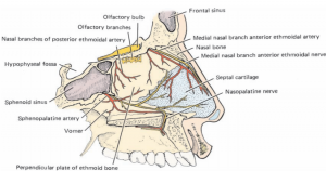
- The medial wall of the nasal fossa—the median nasal septum—is composed of the vomer, the perpendicular plate of the ethmoid, and the median nasal septal cartilage.
- The entire median nasal septum is covered by mucoperiosteum. Frequently, this septum deviates to one side, infringing on the nasal fossa of that side. Associated with the anteroinferior aspect of the septum is the vestigial vomeronasal organ (the Jacobson organ) lying on the vomeronasal cartilage.
- This structure, olfactory in nature, is well-developed in some lower animals.
Lateral Wall
- The lateral wall of the nasal fossa differs from the medial wall. Instead of being relatively smooth, it presents three scrolled laminae of bone that jut medially into the nasal fossa.
- These turbinate bones, covered by mucoperiosteum, are referred to as the superior, middle, and inferior nasal conchae.
- Inferior and lateral, under the cover of the projecting concha, is the correspondingly named meatus. Superior to the superior meatus, just anterior to the body of the sphenoid bone, is the sphenoethmoidal recess containing the ostium (opening) of the sphenoidal sinus.
- A region of another sinus, the posterior ethmoidal air cells, opens below the superior concha into the anterior region of the superior meatus. The middle nasal concha overlies and covers the lateral wall of the middle meatus.
- A marked, rounded projection is formed on this wall by the middle ethmoidal air cells of the ethmoidal air sinus. This rounded projection is known as the ethmoidal bulls inferior to which is a thin, arched ledge of bone, the uncinate process of the ethmoid.
- Located between the bulla and the uncinate process is an arch-shaped opening, the semilunar hiatus, connecting the ethmoidal infundibulum with the middle meatus. The anterior ethmoidal air cells, the maxillary sinus, and, frequently, the frontonasal duct from the frontal sinus open into the ethmoidal infundibulum.
- The inferior nasal concha, usually the largest of the three conchae, is a separate bone, whereas the middle and superior conchae are projections of the ethmoid bone.
- The inferior concha overhangs the inferior meatus, whose inferior extent is formed by the floor of the nasal cavity. The nasolacrimal duct opens into the anterosuperior aspect of the inferior meatus

Floor and Roof
- The floor of the nasal fossa is formed by the horizontal process of the palatine bone and the palatine process of the maxilla.
- The incisive canal, transmitting the nasopalatine nerve and vessels, perforates the mucous membrane of the anteromedial aspect of the floor adjacent to the septum, leading into the incisive foramen.
- The contained nerves and vessels serve the anterior hard palate. The roof of the nasal fossa is concave cranially, and its bony vault is composed of the cribriform plate of the ethmoid bone as well as parts of the sphenoid, palatine, vomer, frontal, and nasal bones.
- The mucous membrane lining the nasal fossa may be classified into two categories: a pink to red, richly vascularized respiratory mucosa lining most of the nasal fossa and moistening the inhaled air, and a yellowish-brown olfactory mucosa, responsible for olfaction, located superiorly.

References
