Feeding patterns
- Macrophagous feeders (eat large food particles) include carnivores which eat meat (includes fish-eating piscivores, and insect-eating insectivores, etc.), herbivores which eat vegetation and omnivores which eat a mixed diet. They often have jaws and teeth.
- Microphagous feeders include suspension feeders (e.g. annelid fanworms or urochordate sea-squirts) which use netting devices to filter and concentrate small particles suspended in water (filter-feeding) and other devices such as mucus strings and parachutes. Fluid feeders include forms such as aphids tapping fluid in plant phloem. Osmotrophs include forms such as tapeworms and other parasites absorbing nutrients through the body surface.
Digestive system Regions
Digestive systems show great variation in the Animal Kingdom. Gut structures do not always correlate with feeding habits (although large stomachs may reflect large, infrequent meals). There is a close correlation between gut structures and the nature of the ingested food: carnivores tend to have short absorptive intestines.
A five-part gut is recognizable.
(1) Reception by the mouth and pharynx. Mechanical trituration can occur here (e.g. use of tongue and teeth in mammals); enzymic digestion may commence; toxic factors may paralyse prey. Fluids lubricate the food.
(2) Conduction and storage in the oesophagus which may be distended to form a crop (birds) or fermentation chambers (ruminant mammals).
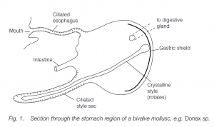
(3) Trituration and early digestion in the stomach and the first part of the intestine. Mechanisms for internal trituration may be found in animals which swallow large food masses [e.g. mammalian stomach muscles; muscles and gravel in bird gizzards; rotation of a crystalline style in the bivalve stomach]. In crustaceans, a chitin-lined gizzard precedes the intestine: food is ground in a gastric mill with dorsal and lateral teeth moved by muscles. Enzymic digestion is commonly found in this region. Diverticula are common (e.g. teleost fishes): cilia may propel food into diverticula in invertebrates (e.g. molluscs).
(4) Absorption and further digestion occurs in the vertebrate small intestine: most of the enzymes come from the pancreas. The liver secretes bile. Some absorption occurs in the duodenum but mostly in the ileum. In most invertebrates, digestion has occurred in stomach diverticula and the intestine is absorptive. The area is increased by typhlosole (longitudinal fold, e.g. earthworm), spiral valve (elasmobranchs), folding and villi (e.g. mammals).
Area of the human intestine:
Simple tube 3.3 × 103 cm2
With folds 104 cm2
With villi 105 cm2
With microvilli 2 × 106 cm2
(5) Compaction and formation of feces where much water is absorbed in terrestrial species (particularly important in insect osmoregulation).
Human gut Structure
- The rather unspecialized human gut reflects our omnivorous diet. The inner surface of the gut is continuous with the exterior of the body so the gut lumen is technically outside of the body.
- The gut is lined by a mucosa (epithelial tissue, underlying basement membrane, connective tissue with thin smooth muscle), a submucosa (outside the mucosa with connective tissue, blood vessels, nerves), muscularis externa (with inner circular muscles and outer longitudinal muscles) and an outer serosa of connective tissue.
- Co-ordinated muscle contractions produce ring-like contractions to churn up food and waves of peristalsis to propel food along the gut. Mouth Food enters the gut via the mouth; mastication by the teeth and the tongue takes place. Saliva, which contains bicarbonate (HCO3–) and an α-amylase, lubricates the bolus of food.
- Approximately 1500 cm3 saliva per day are secreted by the salivary glands; secretion is under parasympathetic nervous control. Human teeth are relatively unspecialized; each jaw quadrant has two biting incisors, one tearing canine, two premolars and three molars for grinding. Teeth are coated with enamel over dentine: in the middle is a vascularized, innervated pulp.
- Teeth are embedded in jaw bones by ligaments and cement. Absorption of some drugs occurs in the mouth (e.g. morphine).
Esophagus
The triturated, lubricated food bolus passes down the esophagus by peristalsis; the esophagus is lined (like the mouth) with stratified squamous epithelium. Peristalsis is movement of a muscular tube, such as the gut, by co-ordinated contractions of longitudinal and circular smooth muscle in a definite (usually anterio-posterior) direction.
Stomach
- Food is triturated further in the stomach by bands of smooth muscle – longitudinal and circular muscles antagonize each other. This allows mixing with gastric juices.
- Food first collects in the relaxed body of the stomach; pronounced peristaltic waves from the top (fundus) to the base (antrum) occur – this generates chyme which is squirted through the pyloric sphincter in small quanta.
- The sphincter is normally open except after a squirt into the duodenum (i.e. it acts as a valve: a large meal can take more than 4 hours to enter the duodenum).
- The stomach mucosa is very thick, with many gastric pits. Mucus-secreting cells cover the stomach surface and line the pits.
- Gastric juice is very acid with hydrochloric acid (HCl, pH = 1.5–2.5), which is secreted by gastric glands in the lower parts of the pits: acid kills bacteria and living cells in the food, loosens fibrous components in food and facilitates conversion of pepsinogen to pepsin.
- Protein digestion is initiated in the stomach: gastric juice contains pepsinogen (an enzyme precursor or zymogen) which is converted to pepsin by gastric HCl.
- Zymogens prevent digestion of the stomach by self-enzymes. Mucus also protects cells from activated enzymes; the mucus forms a protective coat absorbed onto a glycocalyx on the surfaces of microvilli of lining cells.) Rennin precipitates soluble proteins in milk. Water, salts, some vitamins and some drugs (e.g. ethanol) are absorbed in the stomach.
Small intestine
- The small bowel (intestine) comprises, in sequence, the duodenum, jejunum and ileum: it is the site of enzymatic digestion using enzymes from the pancreatic juice and in or on the surfaces of intestinal epithelial cells.
- Pepsin is inactivated by mildly alkaline conditions in the small bowel. Protein digestion continues using pancreatic trypsin and chymotrypsin.
- Pancreatic juice also has an amylase and a lipase. Bile salts from the liver (draining into the duodenum from bile ducts after temporary storage in the gall-bladder; see below) emulsify fats: bile also acts as an excretion medium for hemoglobin degradation products and cholesterol.
- Much of the final breakdown of proteins, fats and sugar polymers occurs within cells lining the small bowel: as intestinal epithelial cells mature and pass up to the tips of the villi, the enzyme contents increase.
- It is now thought that digestion occurs in the cells or on their membranes – there does not seem to be a secretion of large quantities of enzymes into the small bowel lumen (in which those enzymes which are found derive from the pancreas).
- Digestion products are absorbed in the small bowel whose area is increased by villi and microvilli (see above). Some fats, hydolyzed to fatty acids and glycerol, are resynthesized to new fats, and are packaged into chylomicrons and absorbed by lacteals (of the lymphatic system) in the villi: chylomicrons deliver fats to the adipose cells or liver or are broken down in the bloodstream.
- Cholesterol is made in the liver and is packaged into low density lipoprotein (LDL) complexes which are excreted in bile. Stored cholesterol can be repackaged for delivery to cells for membrane or steroid hormone synthesis.
Large intestine
- Absorption of water, sodium and other minerals occurs in the large bowel (intestine). The first part is the colon where, in humans, 7 liters of water are absorbed per day. The colon harbors bacterial flora which can further digest food and synthesize absorbable amino acids and vitamins (e.g. vitamin K).
- The cecum, almost absent in humans, is a blind pouch with the vermiform appendix at its end; it is a secondary lymphoid organ. Residues (dead cells, bacteria, cholesterol, bile pigments, undigested food, especially cellulose fibers) form the feces, stored in the colon. Feces are expelled periodically by passing them down the rectum and out through the anus.
Co-ordination of Digestion
- Enzymes act consecutively along the gut at specific sites: this requires release of small quantities of food and a precise sequence of enzyme release. Food movement is largely under autonomic nervous control. Saliva secretion is nervous alone: it is initiated by food in the mouth, or by the smell, sight or anticipation of food.
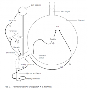
- Gastric juice secretion is initiated by nervous stimulation due to food in the mouth and/or the stomach. Distention of the stomach antrum results in the hormone gastrin being secreted by stomach-lining cells into the bloodstream, stimulating further gastric juice secretion (an isolated, denervated stomach pouch still secretes gastric juice when food is in the antrum).
- Gastrin secretion is inhibited by high acid levels in the stomach (negative feedback). As stomach contents are released into the duodenum, the duodenum secretes a gastrininhibitory polypeptide (GIP) which further inhibits gastric juice secretion.
- Acid stomach contents in the duodenum stimulate release of secretin: this enhances the flow of bicarbonate-rich (alkaline) pancreatic juice. Protein fragments stimulate cholecystekinin–pancreozymin (CCK–PZ) which leads to the release of pancreatic enzymes and stimulates gall-bladder contraction. Motility hormones affect movement of the villi by smooth muscle contraction.
The mammalian Liver
- The liver develops as an outgrowth of the gut; most of its cells are hepatocytes arranged in cylindrical lobules intimately associated with venules, arterioles and bile canaliculi.
- Food-enriched blood is brought from the gut to the liver by the hepatic portal vein, and oxygenated blood is transported to the liver by the hepatic artery.
- The liver is drained by the hepatic vein. Liver cells secrete bile which passes down bile canaliculi into a hepatic duct and then up a cystic duct to the gall-bladder.
- Relaxation of a sphincter at the neck of the gall-bladder and contraction of the bladder following the release of food into the duodenum allows bile to flow down the common bile-duct into the small intestine.
- Bile acts as an excretion medium for cholesterol and hemoglobin degradation pigments, but its main role is to carry bile salts which emulsify fats in the bowel.
- Other functions of the liver include glycogen, amino acid and fat storage and metabolism, detoxification of ammonia to urea, cholesterol synthesis, fetal erythropoiesis (red cell manufacture), breakdown of excess hemoglobin, red blood cell storage, vitamin storage, synthesis of many plasma proteins and heat production
The mammalian Pancreas
Like the liver, the pancreas develops as an outgrowth of the small intestine, its cells producing large quantities of digestive enzymes which travel to the gut via the pancreatic duct, which joins the common bile duct just before its junction with the bowel. The islets of Langerhans are important endocrine glands producing hormones (e.g. insulin, glucagon) associated with blood glucose control
Cellulose digestion by symbionts
- Ruminants, for example cows and sheep, have a gut divided into sections similar to those of humans, except that the esophagus and stomach are greatly modified. They use symbiotic bacteria, yeasts and protoctistans to break the β,1–4 links in cellulose.
- There are four chambers: the rumen, reticulum and omasum are sacculations of the esophagus; the abomasum is the true stomach.
- On being swallowed, food goes to the reticulum (tripe) where it is made into cud balls. The fermenting mass is regurgitated to the mouth for further trituration (chewing the cud). On second swallowing, food passes to the rumen.
- The rumen is rich in anaerobic symbiotic microorganisms (especially bacteria and ciliates) which ferment cellulose to fatty acids, carbon dioxide and methane, and starches to sugars. Fatty acids are absorbed by the glandular epithelium of the rumen, the gases are eructed (‘burped’!).
- The fermented mass then passes to the omasum (psalterium): here it is further triturated, strained and squeezed by strong muscular contractions.
- It then passes to the abomasum where digestive juices start work: bacteria are also digested in the abomasum and are rich sources of nitrogen and vitamins of the B complex. Lagomorphs, for example rabbits, have a large cecum and appendix off the colon.
- This contains bacteria which digest cellulose and produce B vitamins. Lagomorphs re-ingest fecal pellets (coprophagy), giving food a second passage through the gut and permitting the products of microorganism cellulose digestion to be absorbed.
- Hindgut fermenters such as horses have a capacious cecum. Symbiotic microorganisms break down cellulose. Digestion products can pass forward, by reverse peristalsis, for absorption in the small bowel.
Mammal tooth Patterns
Basic (‘primitive’) pattern
The basic pattern is three incisors, one canine, four premolars and three molars on each side, in each of the upper and lower jaws. The dental formula is I 3/3 C 1/1 PM 4/4 M 3/3.
Variations
(1) Insectivora (e.g. shrew): unspecialized teeth, similar to the ‘primitive’ pattern.
(2) Rodents (e.g. squirrel): one incisor, no canines or anterior premolars. Diastema between the incisor and back premolars allows cheeks to be drawn in, closing off the front of the mouth. There is enamel only on the front edge of the incisors: dentine wears away leaving a chisel edge. Incisors grow, as do premolars and molars. A longer length of articulation with the skull allows backward and forward movements. There is a nonrigid join between the mandibles: thus two incisors can move relative to each other, for example when opening nuts. All these variations facilitate gnawing.
(3) Lagomorphs (e.g. rabbit): similar to rodents, but two incisors.
(4) Perissodactyls (e.g. horse): three incisors but no canines; the first premolar is vestigial. There is diastema and the molars and premolars are square. The high-crowned teeth, which grow throughout life, have rough, grinding surfaces.
(5) Artiodactyls (e.g. cow): cows have lost all their upper incisors. They crop grass using lower incisors and horny upper gums. A muscular and protrusible tongue sweeps grass into the mouth. They have stout, elongated high-crowned molars with rough grinding surfaces (enamel, dentine and cementum wear away at different rates, so hardened cementum and enamel form ridges).
(6) Proboscideans (e.g. elephant): the nose and upper lip form a prehensile trunk. One upper incisor grows throughout life to form the tusk (for defense). There are no canines or adult premolars; three molars per jaw, used one after another, develop in a series from front to back. Molars consist of many plate-like ‘cones’ joined by cementum producing a hard working surface with transverse ridges.
(7) Primates (e.g. monkey, ape or human): omnivores with low-crowned, unspecialized teeth. They possess two incisors, one canine, two or three premolars and three molars.
(8) ‘Edentate’ groups (e.g. anteaters, pangolins): they have a tendency to lose teeth in evolution. They possess a long tongue with adhesive saliva.
(9) Carnivores (e.g. cat, seal): land forms (fissipedes) have piercing, cutting incisors, large tearing, slashing canines, cutting and shearing molars and premolars (last upper premolar and first lower molar form shearing carnassials). Articulation allows an up-and-down bite. Marine forms (pinnipedes) have peg teeth which hold prey until it is swallowed (the walrus has tusks for sexual display and, arguably, to dig up molluscs and its flattened molars can crush).
(10) Cetaceans (e.g. whales, dolphins): whale-bone whales (e.g. blue whale) have baleen plates to filter-feed krill; toothed whales (e.g. killer whale) have many peg-like teeth to hold prey until it is swallowed (compare seals).