Introduction
Chemical Composition of Bacterial Cell Wall:
- Bacterial cell walls are about 10-25 nm thick and account for about 20-30% of the dry weight of the cell.
- Chemically the cell wall is composed of a mucopeptide (Peptidoglycan or murein) framework formed by N-acetyl glucosamine and N-acetyl muramic acid molecules alternating in chains, which are cross-linked by peptide chains.
- The cell wall of Gram-positive bacteria has a simpler chemical nature than those of Gram-negative bacteria.
- The cell wall carries bacterial antigens that are important in virulence and immunity.
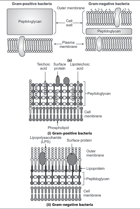
The cell wall of Gram-positive bacteria:
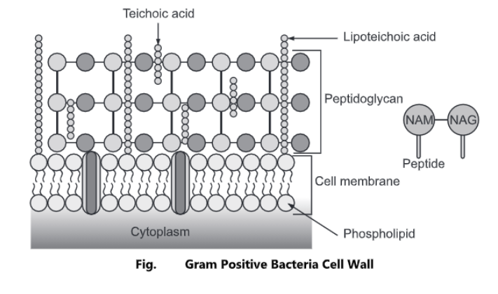
- It is homogenous and thick, about 20 – 80 nm in thickness. It mainly consists of peptidoglycan, teichoic acids, and sometimes other polysaccharides. Peptidoglycan accounts for about 90% of the total dry weight of the cell wall.
- The cell wall of Gram-positive bacteria consists of the following layers:
- Peptidoglycan layer: Peptidoglycan is a complex heteropolysaccharide porous cross-linked polymer that is responsible for the strength of the cell wall. This is thicker (15 – 80 nm) than that of Gram-negative bacteria (2 – 3 nm).
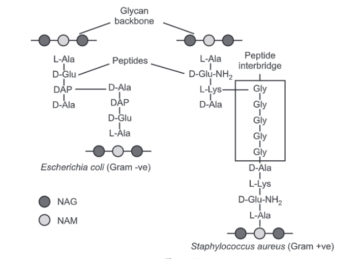
Peptidoglycan is composed of three components:
(a) Glycan backbone: Glycan backbone is the repeated unit of N-acetyl muramic acid (NAM) and N-acetyl glucosamine (NAG) linked by a β-1,4 glycosidic bond.
(b) Tetra-peptide side chain (chain of 4 amino acids) linked to NAM: The glycan backbone is cross-linked by tetra-peptide linkage. The tetra-peptides are only found in NAM. Although the peptidoglycan chemistry varies from organism to organism the glycan backbone i.e. NAG-NAM is the same in all species of bacteria.
The amino acids found in tetra-peptide are:
1. L-alanine: First position in both Gram-positive and Gram-negative bacteria
2. D-glutamic acid: Second position
3. D-aminopimelic acid/ L-lysine: Third position (variation occurs)
4. D-alanine: Fourth position.
(c) Peptide cross-linkage:

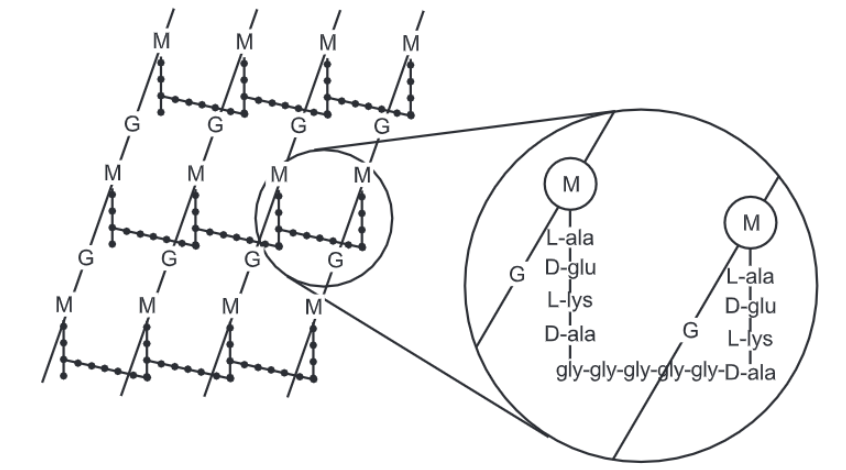
There are many variations in cross-linking of different organisms. Based on this the classes of mureins are as follows:
(i) Group A: It shows the pentaglycine bridge between Meso diaminopimelic acid of one tetrapeptide and D alanine of the other.
(ii) Group B: It shows the pentaglycine bridge between the D glutamate of one tetrapeptide and the D alanine of the other.
(iii) Group C: It shows the pentaglycine bridge between the L lysine of one tetrapeptide and the D alanine of the other.
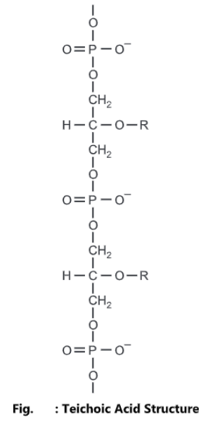
2. Teichoic acid layer: Teichoic acid is a major surface antigen of Gram-positive bacteria. Teichoic acid is a water-soluble polymer of glycerol or ribitol phosphate.
It is present in gram-positive bacteria. It binds to protons and maintains the structure of the cell wall. It binds with metal ions. It acts as a receptor for many viruses. It constitutes about 50% of the dry weight of the cell wall. It is composed of the lipid bilayer, protein, and lipopolysaccharide (LPS) layer.
Two types of teichoic acids are present:
- Cell wall teichoic acid is linked to peptidoglycan.
- Membrane teichoic acid is also known as lipoteichoic acid which is linked to membrane glycolipids.
Examples of Gram-positive bacteria: Micrococcus, Staphylococcus, Streptococcus, and Leuconostoc.
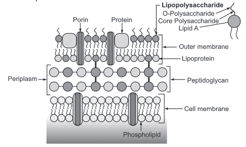
Cell wall of Gram-negative Bacteria:
The cell wall of Gram-negative bacteria is very complex in structure.
It is composed of two layers:
1. Inner layer: It lies next to the cell membrane and is very thin about 2 – 3 nm in dimension. It contains peptidoglycan which accounts for 5 – 10% total dry weight of the cell and forms a gel-like structure. The inner layer is separated from the outer layer by periplasmic space. The periplasm contains a high concentration of degradative enzymes and transport proteins. Gram-negative cell wall does not contain teichoic acid. Due to less percentage of peptidoglycan Gram-negative cells are more susceptible to mechanical damage but less susceptible to lysozyme action.
2. Outer later: The outermost layer of the Gram-negative bacteria cell wall is called the outer membrane, which contains various proteins known as outer membrane proteins (OMP). Porins are one of them which form transmembrane pores that serve as diffusion channels for small molecules. This is a bilayered membrane containing phospholipid molecules facing polar ends outside and non-polar ends inside the layer. It is in contact with peptidoglycan present in the inner membrane through proteins. This layer contains proteins and lipopolysaccharides.
3. Lipopolysaccharides (LPS): LPS are involved in their endotoxin activity and antigenicity. It creates a negative charge in the cell. The LPS consists of three regions:
i. Glycolipid portion (lipid A): This region contains glucosamine sugar. It is embedded in the outer membrane and contains three fatty acid chains and phosphate or pyrophosphate moieties. It is responsible for endotoxic activities like pyrogenicity, lethal effect, tissue necrosis, anticomplementary activity, B cell mitogenicity, immunoadjuvant property, antitumor activity, etc.
ii. Core polysaccharide: It is joined to the lipid A region and contains unusual sugars like 2-Keto-3-deoxy-D-mannooctonic acid (KDO) (a unique eight-carbon sugar), galactose, and n-acetyl glucosamine (NAG).
iii. O-polysaccharide: This portion determines O antigen specificity. It is a short polysaccharide chain extending outside of the core region. It contains galactose, mannose, rhamnose, fucose, xylose, ribose etc. This ‘O’ side chain shows variations in the composition of different organisms and thus is very specific.
Examples of Gram-negative bacteria are E. coli, Salmonella, Klebsiella, Shigella, and Pseudomonas.
Functions of the Cell Wall:
- The cell wall provides definite shape, strength, and rigidity to the cell.
- It also provides protection against mechanical stress and physical shocks.
- It plays an important role in cell division.
- It helps to control cell expansion due to the intake of water.
- It also helps in preventing water loss from the cell.
- It is responsible for transporting substances between and across the cells.
- It acts as a barrier between the interior cellular components and the external environment.
- It protects the cell from toxic substances.
- It enhances pathogenicity by making cells resistant to engulfment by macrophages.