Bacterial Flagella
- It is the locomotory organ in bacteria.
- Bacterial flagella are long, thin (about 20 nm), and whip-like appendages that move the bacteria towards nutrients and other attractants.
- Flagella are free at one end and attached to the cell at the other end.
- Flagellum can never be seen directly with the light microscope but only after staining with special flagella stains that increase their diameter.
- Motile bacteria except for spirochetes possess one or more unbranched, long, sinuous filaments called flagella which are organs of locomotion.
- The filament is external to the cell and connected to the hook at the cell surface. The hook-basal body portion is embedded in the cell envelope. The hook and basal bodies are antigenically different.
- The flagella are 3-20 µm long and are of uniform diameter (0.01-0.013 µm) and terminate in the square tip.
- The wavelength and thickness of the filament are characteristics of each species. Some bacteria exhibit biplicity that is they have flagella of two different wavelengths.
- Flagella are made of a protein known as flagellin.
- The presence or absence of flagella and their number and arrangement are characteristic of different genera of bacteria.
Structure of Flagella:
The structure of bacterial flagella has been described by Simon (1978), Doestsch and Sjoblad (1980), and Ferris and Beveridge (1985).
1. Basal body: It is composed of a central rod inserted into series of rings which is attached to the cytoplasmic membrane and cell wall.
In Gram-negative bacteria, two pairs of rings, the proximal ring, and the distal ring are connected by a central rod. There are two pairs of rings. The outer pair of rings, L-ring and P-ring are attached to the respective polysaccharides and peptidoglycan layer of the cell wall, and the inner pair of rings i.e. S-ring and M-ring are attached to the cell membrane. Four rings are the L-lipopolysaccharide ring, P-peptidoglycan ring, S-super membrane ring, and M-membrane ring.
L-ring: it is the outer ring present only in Gram-negative bacteria, it is anchored in the lipopolysaccharide layer.
P-ring: it is the second ring anchored in the peptidoglycan layer of the cell wall.
M-S rings: anchored in the cytoplasmic membrane.
The outer rings form a bearing for the rod to pass through it. In Gram-positive bacteria only the distal (inner) pair of rings are present. The S-ring is attached to the inside thick layer of peptidoglycan and the M-ring is attached to the cell membrane.
2. Hook: It is the wider region at the base of filament. It connects filament to the motor protein in the base. The length of the hook is longer in gram-positive bacteria than in gram-negative bacteria.
3. Filament: It is a thin hair-like structure that arises from a hook.

There are various types of flagellar arrangements:
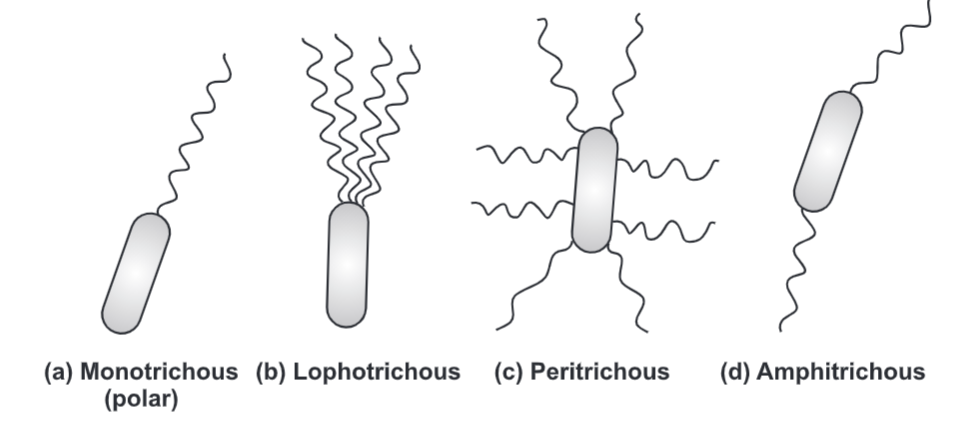
1. Monotrichous: Presence of single flagella in one end of the cell. Examples: Vibrio cholera, Pseudomonas aeruginosa.
2. Lophotrichous: Presence of bundle of flagella in one end of the cell. Example: Pseudomonas fluorescence.
3. Amphitrichous: Presence of single or cluster of flagella at both ends of the cell. Example; Aquaspirillium.
4. Peritrichous: Presence of flagella all over the cell surface. Examples: E. coli, Salmonella, Klebsiella.
5. Atrichous: Absence of flagella. Example: Shigella.
Mechanism of Flagellar Motility:
- The movement of flagella results from the rotation of the basal body which is like the movement of the shaft of an electric motor. The movement of flagella is due to rotational force or ‘Torque’ created by proton motive force. This proton motive force is generated due to the translocation of protons from the ‘M’ ring. This provides a chemical gradient ie. a concentration gradient that shows a higher concentration of protons outside and less concentration inside the cytoplasm. It also creates a pH gradient across the membrane. This chemical gradient provides chemical energy which results in the revolution of flagella. The interaction of S and M ring allows movement of the hook and filament.
- At the base surrounding the inner ring (M-S and C ring), there is a series of proteins called Mot protein. A final set of proteins called Fli protein functions as a motor switch. The flagella motor rotates the filament as a turbine causing movement of the cell in the medium. A turning motion is generated between S-ring and M-ring. S-ring acts as a starter while M-ring acts as a rotor. The basal body as a whole gives a universal joint to the cell and allows complete rotation of hook and filament. The rotation of flagella is either clockwise or anticlockwise.