Cell Coat and its function
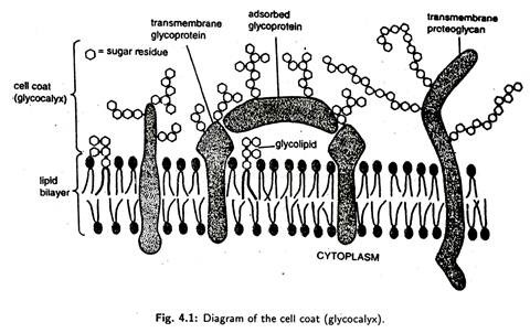
The plasma membrane is surrounded and protected by the cell coat. Sometimes, the cell coat is also called glycocalyx, because it contains sugar units in glycoproteins and polysaccharides. The cell coat is found to be equivalent to the oligosaccharide side chains of glycolipids and glycoproteins that stick out from the cell surface and are covalently attached to protein moieties. However, in many types of cells, there is a separate “fuzzy layer”, beyond the cell coat, which is composed mainly of carbohydrates and is secreted by the cell.
The cell coat can be stained with PAS or Alcian blue for light microscopy and with lanthanum or ruthenium red for electron microscopy. By the use of lectins, the carbohydrates can be specifically observed. Lectins are proteins that are normally derived from the plants and they tend to bind to the cell surface and cause agglutination. They are used as recognition molecules for the sugar components of glycoproteins. Concanavalin A is a lectin that has been isolated from jack beans and is specific for glucose and mannose residues. Another lectin, called germ agglutinin, is specific for N-acetylglucosamine. These lectins may be labelled with fluorescent dyes or with electron-dense materials for electron microscopic observation.
The cell coat is a 10 to 20 nm thick layer and is in direct contact with the outer leaflet of the plasma membrane. In Amoeba, the cell coat is formed by fine filaments — 5 to 8 nm thick and 100 to 200 nm long. Chemically, the cell coat has negatively charged sialic acid termini, on both glycoproteins and gangliosides. This acid tends to bind the ions of Ca2+ and Na+. The strength of the cell coat varies from cell to cell. For example, the cell coat of the intestinal epithelium is quite strong—it resists vigorous mechanical and chemical attacks; in other cells, the coat is labile and may be depleted by washing or enzyme exposure (Luft, 1976).
The cell coat is the secretion product of the cell that is incorporated into the cell surface and undergoes continuous renewal. As already discussed, the glycoproteins of glycocalyx are synthesized on the ribosomes of RER and their final assembly with oligosaccharide moiety is attained in the Golgi apparatus.
Extracellula Materials
In certain cells, outside the cell coat proper and the fuzzy layer exist the extracellular materials, e.g., jelly coat of eggs of fishes and amphibians, the basal laminae of epithelia, the matrix material in which cartilage and bone cells are embedded and the cell wall of plant cells. In these extracellular materials, the most conspicuous components are collagens and mucopolysaccharides (glycosaminoglycans).
Functions of Cell Coat
In addition to the protection of the plasma membrane, the cell coat performs the following important functions :
(i) Filtration. The extraneous coats sometimes act as filters. For instance, the extraneous coats surrounding the blood capillaries of most vertebrates, especially the kidney glomerulus act as filter and regulate the passage of molecules through it. The extracellular coats of connective tissues contain the chemical compound hyaluronate which controls the diffusion.
(ii) Maintenance of the micro-environment of the cell. The extraneous coats of animal cells can affect the concentrations of different substances at the surface of the cell. For example, a muscle cell with its excitable plasma membrane which is surrounded by a glycocalyx is found to maintain the micro-environment of a muscle cell by trapping the sodium ions.
(iii) Enzymes. The cell coat of intestinal microvilli is found to contain a variety of enzymes that are involved in the terminal digestion of carbohydrates and proteins. For example, it contains the enzyme alkaline phosphatase.
(iv) Immunological properties of the extraneous coats. Some substances of extraneous coats provide immunological properties to the cell. For instance, the plasma membrane of mammalian erythrocytes is found to contain some specific, genetically determined substances (carbohydrates and proteins) corresponding to the A, B and O blood groups. The major sialoglycoproteins of the red blood cell membrane carry the M and N antigens that appear infrequently in men. The cell coat also contains the receptor sites for the influenza virus and for various lectins.
(v) Histocompatibility. The cell coats of some cells contain some antigens which provide histocompatibility, i.e., they permit the recognition of the cells of one organism and rejection of other cells that are foreign to it (e.g., the rejection of grafts from another organism).