Connective Tissue
- It is the most abundant and widely distributed tissue system in the body.
- It binds together, supports and strengthens other body tissues as well as protects and insulates internal organs.
- It is made up of fibres, cells and ground substances.
Fibres:
Three types of fibres are embedded in the extracellular matrix between the cells.
These fibres strengthen and support connective tissues.
(a) Collagen fibres:
These are very strong and allow tissue flexibility. These are made up of protein collagen. It is the most abundant protein making up about 25% and 35% of the total body protein. Collagen fibre is often presented in parallel bundles. It is found in bone, cartilage, tendons and ligaments.
(b) Elastic fibres:
These fibres are smaller in diameter. These are made up of protein elastin surrounded by a glycoprotein named fibrillin which gives strength and stability to tissue. Elastic fibres have the ability to return to their original shape, a property called elasticity. These are found in the skin, lungs, arteries, veins, elastic cartilage, periodontal ligament and foetal tissue.
(c) Reticular fibres:
They consist of collagen protein arranged in fine bundles covered with glycoprotein. These are much thinner than collagen fibres. They give support and strength. These are found in the liver, bone marrow and lymphatic organs.
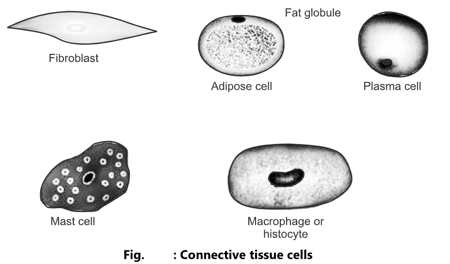
Cells:
Each cell consists of fibroblasts, macrophages, plasma cells, mast cells, adipocytes and white blood cells.
(a) Fibroblasts:
They are the chief cells of connective tissue. They are large, flat cells with branching processes.
(b) Macrophages:
These cells develop from monocytes, a type of white blood cells. There are two types of macrophages. These cells play an important role in the immune response. Fixed macrophages are present in particular tissue such as alveolar macrophages in lungs or spleen macrophages in spleen. Wandering macrophages have the ability to move throughout the tissue and gather at the site of infection to carry phagocytosis.
(c) Plasma cells:
A small cell that develops from a type of white blood cell is called β-lymphocytes. It takes an important part in the immune response. They are present in the gastrointestinal and respiratory tract, salivary glands, lymph nodes, spleen and red bone marrow.
(d) Mast cells:
They produce histamine that dilates the small blood vessels as a part of the inflammatory response.
(e) Adipocytes:
These are also called fat cells or adipose cells. They store fats. They are found deep in the skin and around the heart and kidneys.
(f) White blood cells:
In response to the inflammatory reaction, they migrate from the blood into connective tissue. E.g. Neutrophils gather at sites of infection and eosinophils migrate to the sites of allergic response.
You May Read: Epithelial Tissue: Types, Structure and Function
Ground Substance:
- It is an amorphous gel-like substance present surrounding the cells.
- In the ground substance, cells and fibres are suspended.
- It supports the cells, binds them together, stores water and provides a medium through which substances are exchanged between blood and cells.
- It is primarily composed of water, glycosaminoglycan (hyaluronan), proteoglycans, glycoproteins, hyaluronic acid, chondroitin sulfate and dermatan sulfate.
Functions:
- It acts as an energy store.
- It provides protection to different body organs.
- It provides a structural framework to the body
- It connects different body tissues.
- It connects epithelial tissues to muscle fibres.
- It supplies hormones all over the body.
Classification of Connective Tissue:
Types of Connective tissues are as follows:
Loose Connective Tissue:
- Areolar connective tissue
- Adipose connective tissue
- Reticular connective tissue
Dense Connective Tissue:
- Dense regular connective tissue
- Dense irregular connective tissue
- Elastic connective tissue
Cartilage:
- Hyaline cartilage
- Fibro cartilage
- Elastic cartilage
Bone tissue:
Liquid Connective Tissue:
- Blood tissue
- Lymph
Loose Connective Tissue
- The fibres are loosely woven. It has a large proportion of ground substance. They are easily distorted. On distortion, they become tough and resist further deformation.
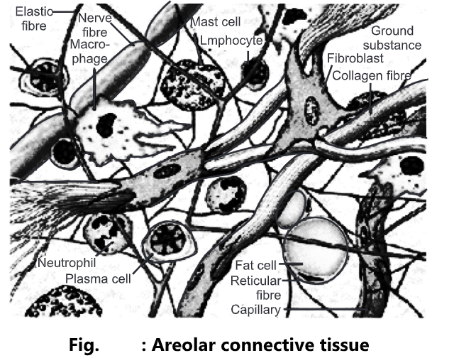
Areolar Connective Tissue:
- They form a loose network in the intercellular material and are not arranged in a particular pattern.
- It consists of collagen fibre, elastic fibres, reticular fibres and several kinds of cells such as fibroblasts, macrophages, plasma cells, adipocytes and mast cells embedded in ground substances.
- Location: It is present below the skin, fill the spaces between muscles, supports blood vessels and nerves in the alimentary canal.
- Yellow elastic fibres are found in arteries and white elastic fibres are found in kidneys and the brain.
- Functions: It gives strength, elasticity and support to tissue.
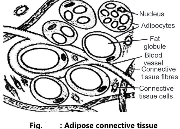
Adipose Connective Tissue:
- It consists of adipocytes that stores fats as a large centrally located droplet.
- Location: It is present in the subcutaneous layer deep in the skin, around the heart and kidneys and yellow bone marrow.
Functions:
- It prevents heat loss from the body.
- It acts as a reservoir of energy.
- It gives shape to the limbs and body
- It protects the underlying organ from injury.
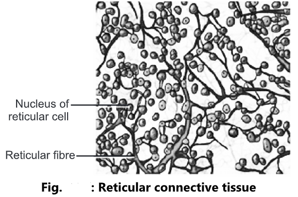
Reticular Connective Tissue:
- It consists of reticular fibres and reticular cells.
- Location: It is present in the supporting framework of the liver, spleen, lymph nodes, red bone marrow and is also found around blood vessels and muscles.
- Functions: It forms the stroma of organs, binds together smooth muscle tissue cells, filters and removes worn-out blood cells in the spleen and microbes in the lymph node.
Dense Connective Tissue
- In this, fibres are densely packed, the fibres content is higher and cell content is lower as compared to loose connective tissue.
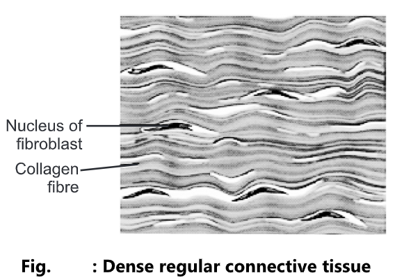
Dense Regular Connective Tissue:
- Bundles of collagen fibres are arranged in parallel patterns to provide strength to the tissue. Fibroblasts appear in rows between the fibres. It is silvery-white in colour and tough in nature.
- Location: It forms tendons (attach muscle to bone) and ligaments (attach bone to bone).
- Functions: It provides a strong attachment to structures.
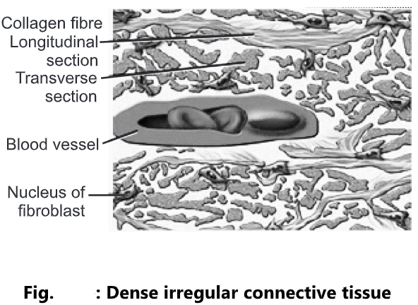
Dense Irregular Connective Tissue:
- It contains collagen fibres that are irregularly arranged and a few fibroblasts appear in rows between the fibres.
- Location: It is present in the tissue beneath the skin, the dermis of the skin, periosteum of bone, membrane capsules around kidneys, liver, testes, lymph node, pericardium of heart and heart valves.
- Functions: It provides strength to different organs.
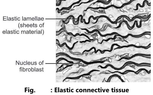
Elastic Connective Tissue:
- It consists of freely branching elastic fibres. Fibroblasts are present in spaces between fibres. It is yellowish in colour.
- Location: It is present in the lung tissues, wall of elastic arteries, trachea, bronchial tubes and vocal cords.
- Function: It allows the stretching of various organs.
Cartilage
It consists of a network of closely packed collagen fibres and elastic fibres embedded in a gelatinous substance called chondroitin sulfate of the ground substance. The cells of mature cartilage are called chondrocytes.
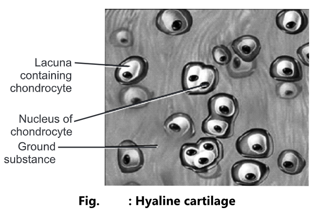
Hyaline Cartilage:
- It is bluish-white in colour. It consists of fine collagen fibres and many chondrocytes. It is the most abundant cartilage in the body.
- Location: It is present at the ends of long bones, anterior ends of ribs, nose, and part of larynx, trachea, bronchi, bronchial tubes, embryonic and fetal skeleton.
- Functions: It provides small surfaces for movement at joints, flexibility and support.
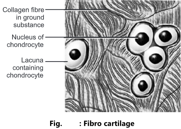
Fibro Cartilage:
- It is the strongest form of cartilage. The chondrocytes are scattered among the bundle of collagen fibres within the extracellular matrix.
- It is tough and slightly flexible.
- Location: It is present in the intervertebral disc.
- Functions: It covers and protects the bony structures of the body.
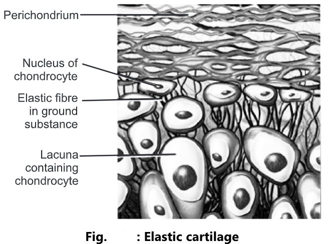
Elastic Cartilage:
- The chondrocytes are located within a threadlike network of elastic fibres within the extracellular matrix.
- Location: It is present in the pinna of the ear and top of the larynx.
- Functions: It provides strength and elasticity and maintains the shape of certain organs, such as the external ear.
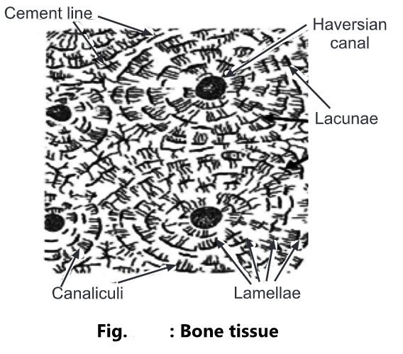
Bone
- It is the hardest connective tissue.
- It has a calcified matrix containing many collagen fibres.
- It is composed of 25% of water, 30% organic material and 45% inorganic salts.
- It is well vascularised.
- It is arranged in concentric ring structures called osteons.
- At the centre of the ring is a structure called as Haversian canal.
- Haversian canal system consists of:
- Central Haversian Channel: It contains blood vessels and nerves.
- Lamellae: Surrounding the central canal, concentric plates of bone are presently called lamellae.
- Lacunae: It contains mature bone cells called osteocytes.
- Canaliculi: Projecting from the lacunae are canaliculi, a network of minute canals containing the processes of osteocytes.
- Location: It is present in compact and spongy bone tissue.
Functions:
- To form a supporting framework for the body.
- To give protection to delicate organs.
- To form joints essential for locomotion of body.
- To form red blood cells in the red bone marrow.
- To provide a store of calcium salts.
- It gives support and maintains shape.
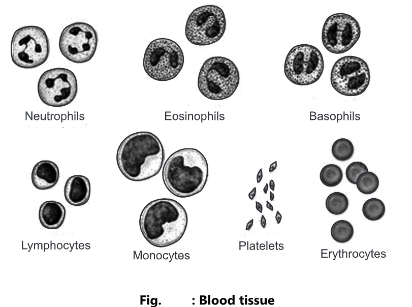
Blood
- It is a connective tissue with a liquid extracellular matrix called blood plasma.
- The blood cells are suspended in the blood plasma.
- It is composed of 55% plasma and 45% of cells.
- Blood plasma is a straw-coloured liquid in which the blood cells are suspended.
- Plasma is composed of 90-92% of water, 7% plasma proteins and clotting factors, and 1% of mineral salts, sugar, hormones and vitamins.
- Blood cells are of three types;
- Erythrocytes (RBC): These cells transport oxygen to body cells and remove carbon dioxide from them.
- Leucocytes (WBC): These cells are involved in phagocytosis, immunity and allergic reaction.
- Thrombocytes (Platelets): These cells participate in the process of blood clotting.
- Location: It is present within blood vessels (arteries, arterioles, capillaries, venules and veins) and within the chambers of the heart.
Functions:
- RBCs transport oxygen to body cells and remove carbon dioxide from them.
- WBCs are involved in phagocytosis, immunity and allergic reaction.
- Platelets participate in the blood clotting process.