Experiment 1
Aim: Cultivation of viruses in the cell line method.
Introduction
Viruses are acellular pathogenic organisms that rely on the host cells for their multiplication and growth because of the lack of their own metabolic machinery. Now a day’s monolayer cell cultures are most widely used in diagnostic and research purposes in viral diseases. Cell culture technique can be performed with the help of chick embryo fibroblast cells, human amnion cells, etc.
The cell lines are classified into three types
- Primary cell line
- Secondary cell line
- Continuous cell lines.
Principle
Viruses are not able to grow on culture media as that of bacteria and they require healthy living cells called host cells. Living cells support the replication of viruses from which viruses can be harvested for further tests. Cells obtained from multicellular eukaryotes, especially animal cells are most preferable.
The growth of the viruses in the cell line can be detected by changes in the morphology of infected cells. Cells may get killed by the infection of viruses. These changes are collectively called as cytopathic effect. But some viruses do not show any cytopathic effect.
Requirements
- Reagents and Media: Minimum essential media (MEM), sodium bicarbonate, EDTA trypsin mixture, fetal calf serum, sterile double distilled water, spirit, sodium hypochlorite.
- Equipments: Inverted Microscope, incubator, haemocytometer, biological safety cabinet
- Apparatus: Sterile glasswares, pre-sterilized tissue culture, plastic wares.
- Specimen Sample: Virus sample from CSF, Stool, Rectal swab, Throat swab
Procedure
- Form the monolayer of cell culture in a sterile flask which is dispersed with the treatment of trypsin and versene.
- Discard the trypsin and versene mixture from the flask.
- Add a small amount of MEM and 10% FCS to the monolayer of the cell.
- Count the number of monolayer cells in a haemocytometer.
- Inoculate the cells with the viral suspension by using the Pasteur pipette in the flask.
- Fill the new flask with MEM and incubate in the horizontal position.
- Incubate the flask at 37°C.
- Select a layer of cells that are infected with viruses and which show morphological changes.
- Observe the cytopathic changes in the monolayer of cells during 7 days of incubation.
Observation: After incubation, examine the flask of the healthy monolayer of cells and as well as infected cells. The morphological changes that occurred in cells are linked with the multiplication of viruses.
Result: Cells showing any cytopathic changes is an indication of multiplication of viruses and vice-versa.
Application
- Cell culture is the most convenient and advanced technique used for the cultivation of viruses.
- Cell culture technique is used for isolation of viruses from the clinical specimens and for diagnosis of viral diseases.
- Biochemical studies like the replication of viruses are also studied by the cell line method.
- Number of viruses are used for the production of Vaccines against different viral diseases.
Drawback
- Not all types of viruses are grown by this cell line method.
- This technique is not an efficient method for the cultivation of viruses.
Key points:
- Non-Cytopathic cells are identified by different methods like Immunofluroscence Haemagglutination, Haemadsorption, and adsorption methods.
- Susceptible cells should be selected for the cultivation of viruses.
Experiment 2
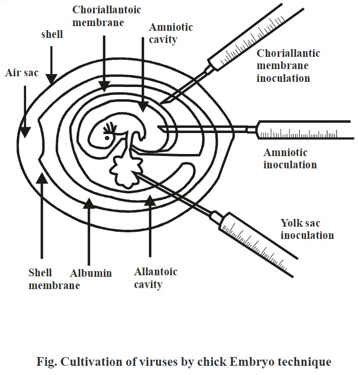
Aim: Cultivation of viruses by Embryonated egg method.
Introduction
The Chick embryo technique is the first technique used for the isolation of viruses. Bulk preparation of the viral suspension is required for the production of vaccines which can be obtained with the help of the chick embryo technique.
Advantages of chick embryo:
- Eggs are easily available and inexpensive.
- Different sites are available in the egg which provides site for the cultivation of viruses.
- Egg lacks their own defense mechanism and hence do not interfere with the cultivation of viruses.
Certain viruses get fail to grow on primary inoculation into eggs.
Principle
Usually, an 8-12 day old chick embryo is preferred. Viruses are introduced in the egg by different routes like yolk sac, amniotic sac, allantoic cavity, or chorioallantoic membrane. Inoculated egg is incubated for 2-10 days. The growth of viruses is indicated by death of embryo and/or formation of pocks. Different sites of egg embryo are use for cultivation of particular virus. Amniotic and allantoic cavity is used for primary isolation of influenza. Yolk sac is used for growth of viruses as well as parasite. CAM is mainly required for pox virus.
Requirements
- Host: Chick embryo
- Specimen: Isolated Viruses sample
- Chemicals: melted paraffin wax, 70% ethanol
- Equipment: Candling lamp, hole puncher
- Apparatus: Disposable Syringes, Gloves, sterile forceps, Pasteur pipette, sterile vials
Procedure
- Check the position of the embryo in the egg and its viability.
- Disinfect the egg with ethanol and remove any contaminants from the shell.
- Drill the egg shell and form very minute hole.
- Inoculate the viral specimen inside the specific site of embryo seal it with melted wax.
- Incubate for 2-10 days and observe.
Observation
Examine the inoculated portion of egg and observe any changes occurred due to viral infection and cultivation.
Result: Viral cultivation is identified by death of embryo.
Application
- Embryonated eggs are useful for the cultivation of avian viruses, and influenza viruses.
- Used in bulk production of vaccines.
Key points
- Eggs should be first candled to determine the position of the embryo and its viability.
- The specific membrane should be chosen for the inoculation of that type of virus.
- Aseptic conditions should be maintained throughout the procedure.