Introduction
- The lymphatic system is a major part of the body’s immune system.
- The lymphatic system is a subset of the circulatory system, with a number of actions.
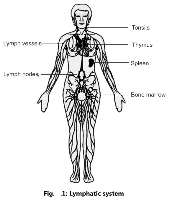
- The lymphatic system is a network of organs, lymph nodes, lymph ducts, and lymph vessels that make and move lymph from tissues of the bloodstream.
- A lymphatic system is a specialized form of reticular connective tissue that consists of tissues and organs that produce, mature, and store lymphocytes and macrophages, for the body’s defense purposes.
- It acts as a transport channel that carries white blood cells to and from the lymph nodes into the bones and antigen-presenting cells to the lymph nodes.
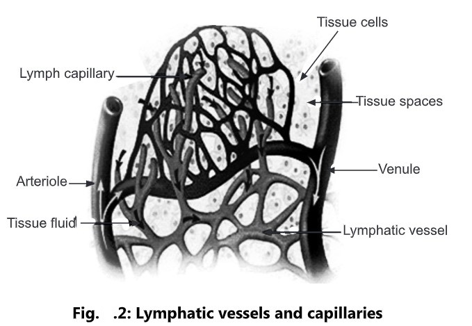
- Lymphatic capillaries reabsorb excessive tissue fluid and transport the fluid through the lymphatic pathway and ultimately dispose of it into the blood.
- Lymphatic vessels carry lipid and lipid-soluble vitamins absorbed by the gastrointestinal tract to blood.
Parts of the Lymphatic system
- Lymph
- Lymphatic vessels
- Lymph trunks and ducts
- Thoracic (Left lymphatic) duct
- Right lymphatic duct
- Lymphatic tissue
- Lymph nodes
- Tonsils
- Spleen
- Thymus gland
Lymph
- The excess interstitial fluid which drains into the lymphatic capillaries is called as lymph.
- It is a clear watery fluid, similar in composition to plasma, with the exception of plasma proteins.
- Lymph transports the plasma proteins that seep out of the capillary beds back to the bloodstream.
- It also carries away bacteria and cell debris from damaged tissues, which are then filtered out and destroyed by the lymph nodes.
- Lymph contains lymphocytes, which circulate in the lymphatic system allowing them to patrol the different regions of the body.
- In the small intestine, fats absorbed from the lymphatics capillaries called as lacteals give the lymph, a milky appearance.
Chemical composition:
- Proteins (g/100 ml): 2.6
- Chloride (m.eq/lit): 116
- Calcium (m.eq/lit): 4.6
- Urea (mg/100 ml): 23.5
Flow of Lymph:
- The sequence of lymph flow:
Blood capillaries (blood)
↓
Interstitial spaces (interstitial fluid)
↓
Lymphatic capillaries (lymph)
↓
Lymphatic vessels (lymph)
↓
Lymphatic ducts (lymph)
↓
Junction of the internal jugular and subclavian veins (blood)
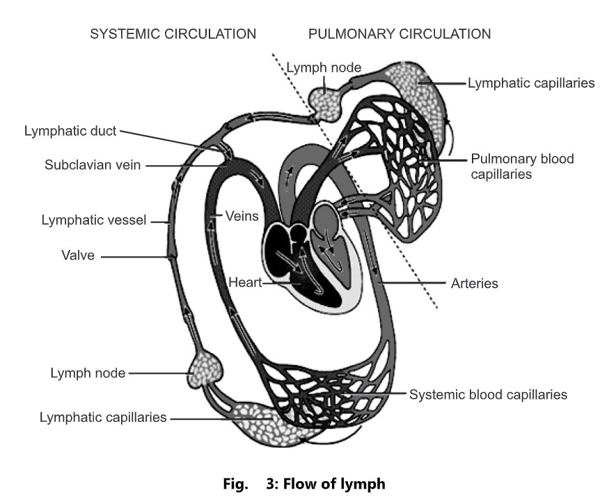
- The lymphatic flow is regulated by means of movements of skeletal muscles and through breathing movements.
- This movement compresses the lymphatic vessels and forces the lymph flow towards the subclavian veins.
- Lymphatic vessels contain a one-way valve that prevents the backflow of lymph.
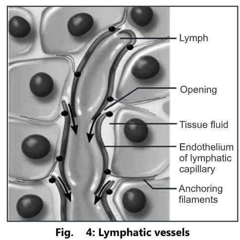
Lymphatic Vessel
- These are tiny thin-walled vessels.
- These are closed at one end.
- The main purpose is to drain the excess of interstitial fluid from around the cell to the venous circulation.
- The wall of lymphatic capillaries is made up of endothelium.
- These are larger in diameter.
- The anchoring filaments hold the endothelial cells to the nearby tissues.
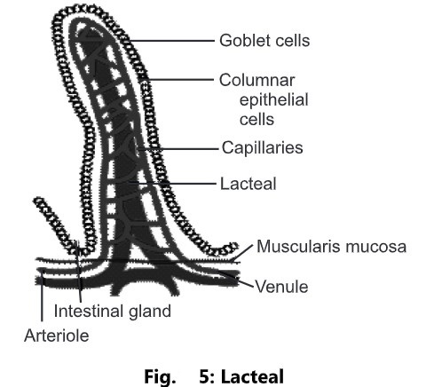
- A lacteal is a lymphatic capillary present in the mucosa of the small intestine
- It absorbs dietary fats and lipid-soluble vitamins from the small intestine.
- A special type of lymph, known as chyle, is produced in the digestive system as lymph absorbs triglycerides from the intestinal villi.
- The chyle has a milky while coloration due to the presence of triglycerides.
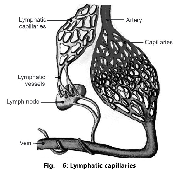
Lymphatic Capillaries
- Lymphatic capillaries combine together to form lymphatic vessels.
- These are thin-walled structures that carry lymph.
- Lymph vessels are lined by endothelial cells.
- A lymph vessel pushes lymph from lymph capillaries to the lymphatic trunk and ducts.
- Lymphatic vessels resemble small veins.
Lymph Nodes
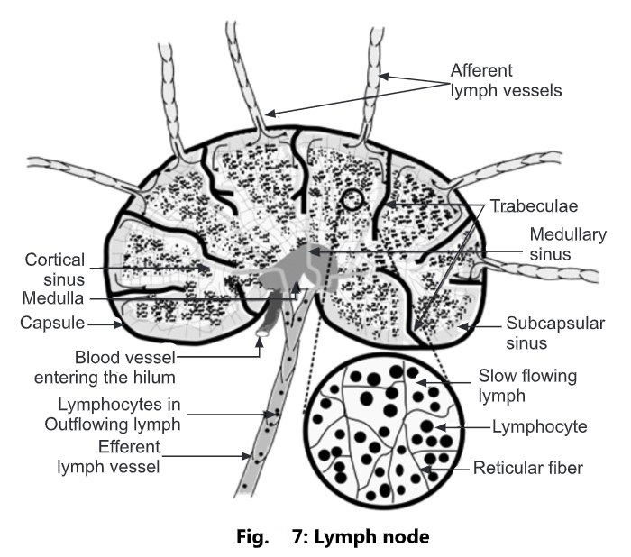
- The oval or bean-shaped organs located along the length of lymphatic vessels are called as lymph nodes.
- They range from 1 to 25 mm in length.
- They are greyish pink in color.
- These are scattered throughout the body, usually in groups.
- These groups are arranged in two sets; superficial and deep.
- Each node is covered by a dense connective tissue called as capsule.
- The capsular extensions are called as trabeculae.
- Internally node has two parts: the outer cortex and the inner medulla.
- The outer cortex contains densely packed lymphocytes arranged in masses called as follicles.
- The outer rim of each follicle contains T-lymphocytes and macrophages.
- In the medulla, the lymphocytes are arranged in strands called as medullary rays.
- Internal to the capsule is a supporting network of reticular fibres and fibroblasts.
- Along with capsule, trabeculae, reticular fibres and fibroblasts constitute the stroma of the lymph node.
- Each node has a concave surface called as hilum.
- Four or five afferent lymph vessels may enter a lymph node while only one efferent vessel carries lymph away from the node.
Functions:
- The lymph node filters foreign substances from lymph as it moves back to the cardiovascular system.
- These substances along with microbes are trapped by the reticular fibres within the node.
- Then lymphocytes and macrophages destroy the foreign substance by phagocytosis.
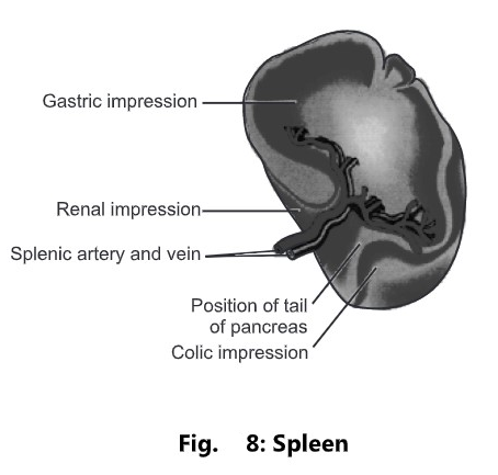
Spleen
- It is a flattened oval organ located in the upper part of the abdomen, under the diaphragm, and behind the stomach.
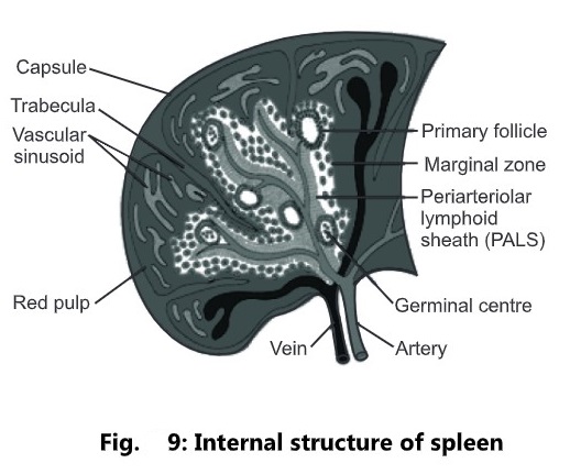
- It is covered by dense connective tissue called a capsule.
- The capsular extensions are called trabeculae.
- Internal to the capsule is a supporting network of reticular fibres and fibroblast.
- Along with capsule, trabeculae, reticular fibres, and fibroblast form the stroma (supporting network) of the spleen.
- The spleen consists of two different kinds of tissue:
- White pulp: It consists of masses of lymphocytes and macrophages
- Red pulp: It consists of blood sinuses.
- Spleen has a concave surface called as hilum.
- The structure entering and leaving the spleen at the hilum are;
- Splenic artery
- Splenic vein
- Lymph vessels
- Nerves
Functions:
- It plays an important role in the phagocytosis of bacteria, damaged RBC’s and platelets.
- During early fetal devolvement, the spleen participates in the blood cell formation.
- Spleen stores and releases blood in times of demand such as during hemorrhage.
- The spleen contains T and B-lymphocytes which are activated by the presence of antigen to fight off infection.