Protoplasm
The cessation of visible activities is due to the cessation of activities within the substance of the body. This living substance’ is known as protoplasm. As long as protoplasm is able to carry on its activities, it is alive; when these activities cease, it is no longer alive. Therefore, life may be studied in terms of the activities of protoplasm.
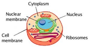
Physical organization of protoplasm
- The amoeba, affords an excellent opportunity to make actual observations on naked living protoplasm.
- A few bodies can be seen in living protoplasm, but most of the structures are practically colourless.
- This makes it necessary to treat it with dyes which stain certain parts. Many different dyes have been employed and numerous methods have been devised for the study of this.
- While most of these result in the death of the protoplasm, the structure is probably not changed very much.
- When examined with a microscope, protoplasm usually looks like a greyish jelly in which may be embedded granules and globules of various sizes and shapes.
- It differs under various conditions; usually, it is about the consistency of glycerin, somewhat viscous but capable of flowing.
- It may exist as a sol that streams easily, or as a more solid gel; under certain conditions, it may change from a sol to a gel or a gel to sol, and back again; this is the unique property of a colloid. Many minute granules can be seen in protoplasm with the aid of a microscope.
- When the protoplasm is in a liquid or sol state, the granules may be observed moving about. This is known as Brownian movement, having been discovered by an English botanist, Robert Brown, in 1827.
- This type of movement is due to invisible particles striking against larger granules. It also occurs in water and other liquidS’ and is not necessarily a sign of life.
- Certain knowledge of the fine structure of protoplasm has been contributed by the physical chemists. They tell us that protoplasm is a colloid and all life is associated with the colloid state.
- Many of the properties of protoplasm depend on the fact that it is a colloid, a mixture in which comparatively large but still invisible particles are suspended in a liquid medium, that is, they do not settle out.
- The particles are estimated to range in size from 0.0001 to 0.000001 mm. in diameter. Colloid suspensions often have a sticky, gluelike consistency; this accounts for the name, which comes from a Greek word that means glue.
- Changes in protoplasm from a sol to the gel condition and back again may be explained on the basis of the distribution of the colloid particles.
- If the particles are more or less evenly distributed in a liquid medium, the enclosed by the meshes, the mixture does not flow but is in a solid or semisolid gel condition.
- Colloidal suspensions and the sol and gel conditions are not confined to protoplasm; for example, jello forms a colloidal suspension in water-when warm it is in a fluid sol condition, but when cool it changes to a solid or semisolid gel condition.
- As in protoplasm, either condition may be changed back into the other. Some other colloidal substances are mayonnaise, cream, butter, glue, and soap mixture flows easily and is in the sol state, but if the particles are arranged so as to form a meshwork, with the liquid medium enclosed by the meshes, the mixture does not flow but is in a solid or semisolid gel condition.
- Colloidal suspensions and the sol and gel conditions are not confined to protoplasm; for example, jello forms a colloidal suspension in water-when warm it is in a fluid sol condition, but when cool it changes to a solid or semisolid gel condition.
- As in protoplasm, either condition may be changed back into the other. Some other colloidal substances are mayonnaise, cream, butter, glue, and soap.
- Consideration of all the known details of the fine structure of protoplasm goes beyond Electron microscope. It uses a beam of electrons and magnetic fields, which take the place of light and lenses. Magnifications of over 100,000 tim~s (diameters) may be obtained.
- This microscope is useful in studying the smallest living things such as viruses and the submicroscopic structures of cells.
- However, the electron microscope reveals that it is far, far more complex than the studies made with the light microscope led us to suspect.
- Life now appears to result from the complex interrelations of the microscopic and ultramicroscopic components of protoplasm.