Transmission Electron Microscope
It is a microscope that uses a beam of accelerated electrons as a source of illumination. As the wavelength of an electron can be 100,000 times shorter than that of visible light photons, electron microscopes have a higher resolving power than light microscopes and can reveal the structure of smaller objects like viruses. Ernst Ruska developed the first electron microscope, a TEM, with the assistance of Max Knolls in 1931. In 1934 Ruska developed an electron microscope that could magnify objects up to 12,000 times. Both light and electron microscopes have the same purpose of producing a magnified image of the object. In a TEM, electrons replace visible light, electromagnetic coils replace glass lenses, and images are viewed on a screen rather than through an eyepiece. The electron beam is accelerated by an anode with respect to the cathode, focused by electrostatic and electromagnetic lenses, and transmitted through the specimen that is in part transparent to electrons and in part scatters them out of the beam. When it emerges from the specimen, the electron beam carries information about the structure of the specimen that is magnified by the objective lens system of the microscope.
Properties of Electrons
- Electrons are used as a source of illumination.
- They are negatively charged subatomic particles.
- When atoms of metal (tungsten) are excited by energy (current of high voltage), the electron velocity is increased to a rate at which electrons leave the orbit, enter the space, and are lost to the atom.
- Electrons emitted from electron guns are passed through an aperture to form a well-defined beam.
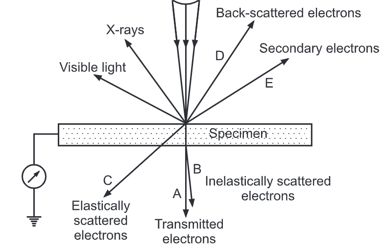
- Electrons interact with the atoms of the biological specimen to form the image.
- The transmitted electrons form the image while the rest of the electrons get scattered.
Components of TEM:
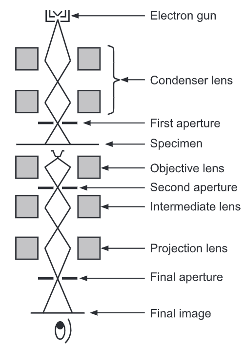
- An electron source: The source of electrons is the electron gun and is located at the top of the microscope body. It consists of a hot tungsten filament cathode as the electron source forming the beam.
- Microscope column: It consists of an evacuated metal tube. It contains a number of electromagnetic lenses, viewing screens, and a photographic plate. The microscope column provides shielding to the operator from X rays that are generated when the electrons strike the metal surfaces.
- Electromagnetic lenses or coils: The Electromagnetic coils correspond to the condenser, objective, and projector coils. Each coil has coils of electric wire wound on the hollow metal cylinder. Electric current passing through the magnetic coil produces an axially symmetric magnetic field in the center of the lens. The electron beam passes through the microscope column and gets deflected by a variable degree depending on the current flowing through the coil of the lens.
- Vacuum chamber: TEM requires a vacuum inside the microscope column. This is maintained with the help of high vacuum pumping and every section of the microscope is kept under high vacuum.
Condensers
Sample stage:
- Phosphor or fluorescent screen: As the electrons are harmful to our eyes the magnified image is observed on a fluorescent screen coated with a phosphor or scintillator material such as zinc sulfide.
- Computer: The image can be photographically recorded by exposing a photographic film or plate directly to the electron beam, or a high-resolution phosphor is coupled by means of a lens optical system to the sensor of a digital camera. The image detected by the digital camera is displayed on a monitor of the computer.
You May Read: Scanning electron microscope
Working of TEM: Image Formation
A Transmission Electron Microscope produces a high-resolution, black and white image from the interaction that takes place between prepared samples and energetic electrons in the vacuum chamber. Image formation occurs by electron scattering. Electrons strike the atomic nuclei and get dispersed and form the image. The electron image is converted to a visible form by projecting it on the fluorescent screen.
- The electrons in the form of a beam pass through a condenser coil and fall on the object.
- They get scattered and transmitted through the object and pass through the objective coil which magnifies the image of the object.
- The projector coil further magnifies the image and projects it on the fluorescent screen.
- The image formation occurs when the energy of the electrons is transformed into visible light.
- Those electrons which reach the fluorescent screen form the bright spots while the areas where electrons do not reach the fluorescent screen form the dark spots.
- The varying degree of the intensity of electrons forms the image with varying degrees of grey. The lighter areas of the image represent the places where a greater number of electrons were able to pass through the sample and the darker areas reflect the dense areas of the object. These differences provide information on the structure, texture, shape, and size of the sample.
- Electron dispersion is due to atomic nuclei containing protons and neutrons.
- The higher the atomic number, the greater the dispersion. Biological materials have low atomic numbers, so the dispersion of electrons is poor. This results in very poor contrast in the image formation.
- In order to increase the contrast number of salts with high atomic numbers is used.
- In order to have maximum magnification, an intermediate coil is fitted between the objective and the projector coils. If the magnification of the objective is 100 and the projector coil is 200, the total magnification becomes 20,000. This can be increased up to 1,60,000 by fitting the intermediate coil. the new generation of hardware correctors can reduce spherical aberration to increase the resolution in high-resolution transmission electron microscopy (HRTEM) to below 0.5 angstroms (50 picometres), enabling magnifications above 50 million times.
- During transmission, the speed of electrons directly correlates to electron wavelength; the faster electrons move, the shorter wavelength and the greater the quality and higher resolution of the image.
- Electron microscopes are used to examine the ultrastructure of a wide range of biological and inorganic specimens including microorganisms, cells, large molecules, biopsy samples, metals, and crystals. Modern electron microscopes produce electron micrographs using specialized digital cameras to capture the images.
- Biological tissue specimens are chemically fixed, dehydrated, and embedded in a polymer resin to allow ultrathin sectioning. Sections of biological specimens, organic polymers, and similar materials may require staining with heavy atom labels in order to achieve the required image contrast.
Advantages:
- TEMs offer the most powerful magnification, potentially over one million times or more.
- A Transmission Electron Microscope is ideal for several different fields such as life sciences, nanotechnology, medical, biological, and material research, forensic analysis, geology, and metallurgy as well as industry and education. TEMs provide information on the element and compound structure.
- Images are high-quality and detailed.
- TEMs provide topographical, morphological, compositional, and crystalline information. The images allow researchers to view samples on a molecular level, making it possible to analyze structure and texture.
Disadvantages:
- TEMs are large and very expensive.
- Laborious sample preparation.
- The need for extremely thin sections of the specimens, typically about 100 nanometres.
- Samples are limited to those that are electron transparent, able to tolerate the vacuum chamber, and small enough to fit in the chamber.
- TEMs require special housing and maintenance.
- Images are black and white.
- Electron microscopes are sensitive to vibration.
- A Transmission Electron Microscope requires constant upkeep including maintaining voltage, currents to the electromagnetic coils, and cooling water.
Techniques Used in Electron Microscopy
Negative Staining:
It is a simple method of studying the morphology and structure of viruses, cell components like ribosomes, cell membrane as well as macromolecules like proteins. The shapes can be determined at the molecular level. The specimen is embedded in a negative stain- a metal in its salt form (potassium phosphotungstate). After drying the electron-dense metal ions surround the specimen. The difference between the specimen and the surrounding heavy metal ions with respect to density produces the contrast. The bright specimen is observed in the dark background of dried stain. The electrons pass through the specimen but not through metallic background. There is no reaction between the stain and the specimen. It is accomplished by using the stain at a pH at which there is no or negligible attraction between the specimen and the stain
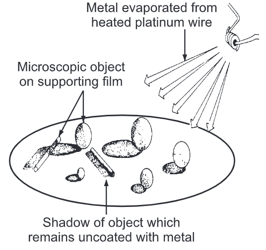
Shadow Casting:
This technique is used to increase the contrast. The specimen is dried on a special grid and placed in a vacuum jar. A heavy metal like chromium, palladium, platinum, or uranium is evaporated at an angle from a filament of glowing tungsten. As it gets deposited at an angle, it piles up on the side from which is deposited while the other side remains clear of it. Under TEM the sides with deposition show dark areas while the other side shows bright shadows. We can get information about the shape of viruses and bacteriophages. It can determine the height of the object. It can give a three-dimensional picture that can’t be obtained by other methods.
Difference Between Light microscope and Transmission electron microscope
Character | Light microscope | TEM |
Illumination | Visible light | Electron beam |
Source of illumination | Sunlight or electric bulb | Electron gun |
Medium of travel | Air | High vacuum |
Mounting of the specimen | Glass slide | Metal grid |
Mechanism of focussing | Adjustment of position of glass lenses mechanically | Adjusting current to the magnetic lens |
Image formation | The collective effect of objective and eyepiece lenses | The combined effect of magnetic condenser and projector lenses |
Source of contrast | Differential light absorption | Scattering of electrons |
Image visualization | With eye | Fluorescent screen |
Maximum magnification | 1000X to 1500X | More than 100,000X |
| Highest resolution | 0.2 pm | 0.5 nm |
References: