Veins of the Neck
The veins of the neck include the external jugular, internal jugular, vertebral, and subclavian veins
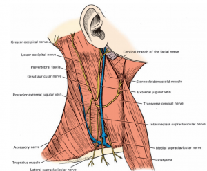
External Jugular Vein
- The external jugular vein is formed by the union of the posterior auricular and retromandibular veins just posterior to the angle of the mandible, sometimes within the body of the parotid gland.
- It passes straight down the neck, under the cover of the platysma muscle and associated superficial fascia, superficial to the fleshy belly of the sternocleidomastoid muscle.
- Along its path it crosses this muscle at an oblique angle.
- Once it reaches the subclavian triangle, the external jugular vein pierces the investing fascia, parallels the posterior border of the sternocleidomastoid muscle, and dives deep to the clavicle to deliver its blood into the subclavian vein, which it joins.
- The external jugular vein has two pairs of incompetent valves just before it empties into the subclavian vein.
- Several tributaries join the external jugular vein, namely, the posterior external jugular vein, which drains the superficial aspect of the back of the neck, and two others, the transverse cervical and suprascapular veins.
- The last two veins drain the region of the shoulder. Another superficial vessel, the anterior jugular vein, occasionally empties into the external jugular vein, but usually it joins the subclavian vein directly.
- The anterior jugular vein is variable, but normally it begins at the level of the body of the hyoid bone and descends parallel to the anterior midline of the neck.
- Inferiorly, near the origin of the medial head of the sternocleidomastoid muscle, the anterior jugular vein pierces the superficial lamina of the investing layer and turns laterally, pierces the posterior lamina, and joins the subclavian (or occasionally, the external jugular) vein.
- While it is between the two laminae of the investing facia, the anterior jugular vein communicates with its corresponding vein of the other side via a venous connection, the jugular arch, which occupies the suprasternal space.
- The external jugular, posterior external jugular, and anterior external jugular veins have numerous smaller named and unnamed tributaries, which drain the areas in their immediate vicinity
Internal Jugular Vein
- The internal jugular vein is the principal vessel responsible for collecting blood from the brain, superficial aspects of the face, and neck.
- The vessel extends from its dilated origin, the superior jugular bulb housed in the jugular foramen, to its inferior dilation, the inferior jugular bulb terminating in the brachiocephalic vein.
- The internal jugular vein is enclosed in the carotid sheath as it travels the length of the neck, and its tributaries pierce this fascia to deliver their blood to the vessel.
- The internal jugular vein receives blood from the following tributaries: dural venous sinus drainage from with the cranium; the facial vein from the superficial face; the lingual vein from the tongue and floor of the mouth; and pharyngeal, superior and middle thyroid, and, occasionally the occipital veins from the neck.
- The dural venous sinuses and their drainage into the superior bulb of the internal jugular vein.The lingual vein receives several tributaries that drain the tongue and floor of the mouth—the sublingual, dorsal lingual, and deep lingual veins, which follow the paths of their corresponding arteries.
- The small phryngeal veins communicate with the pharyngeal plexus of veins and sometimes deliver their blood to the superior thyroid, lingual, or facial veins instead of to the internal jugular vein.
- The superior and middle thyroid veins both drain the thyroid gland and join the internal jugular vein at its superior and inferior aspects, respectively. Both vessels receive smaller named and unnamed tributaries. The inferior thyroid vein usually delivers its blood into the brachiocephalic trunk.
Vertebral Veins
- Unlike their arterial counterpart, the vertebral veins do not traverse the foramen magnum; instead, they are formed from the confluence of many small tributaries within the suboccipital triangle.
- The vertebral veins enter the foramen transversarium of the axis and form a plexus of veins surrounding the vertebral artery, and descend with it within the foramina transversaria of the remaining cervical vertebrae except the last.
- They end in the brachiocephalic vein or occasionally in the subclavian vein. Tributaries of the vertebral veins include the anterior and accessory vertebral veins and the deep cervical vein.
Subclavian Vein
- The subclavian vein is short because it is the continuation of the axillary vein, and it joins the internal jugular vein to form the large brachiocephalic vein.
- Thus, the subclavian vein extends from the external border of the first rib to the junction with the internal jugular vein, passing anterior to the anterior scalene muscle, which separates it from the subclavian artery.
- Here it lies in front of the subclavius muscle, which acts as a cushion, protecting the underlying vessels and nerves.
- The main tributary of the subclavian vein is the external jugular vein, although frequently the subclavian may receive the dorsal scapular and anterior jugular veins.
- The left subclavian vein receives lymph from most of the body via the thoracic duct, whereas lymph from the right upper quadrant of the body is delivered to the right subclavian vein by the right lymphatic duct.
- These ducts pierce the superior aspects of the subclavian veins, just before these are joined by the internal jugular veins.