Embryology: Definition and Concept
Definition:
- Embryology is a branch of science that is related to the formation, growth, and development of the embryo.
- It deals with the prenatal stage of development beginning from the formation of gametes, fertilization, formation of the zygote, development of embryo and fetus to the birth of a new individual.
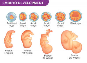
- The ball of dividing cells that result after fertilization is termed an “embryo” for eight weeks and from nine weeks after fertilization, the term used is “fetus.”
- Once an egg is released from the ovary during ovulation, it meets with a sperm cell that was carried to it via the semen.
- These two gametes combine to form a zygote and this process is called fertilization. The zygote then begins to divide and
- The organism is first one cell (the zygote), which then divides, and this process is repeated over and over again. It becomes a blastula Initially these cells are undifferentiated: they have the potential to form any part of the developing body (i.e. pluripotent stem cells).
- The blastula continues to develop, eventually forming a structure called the gastrula. However, over time, cells become progressively more differentiated. They acquire particular characteristics of the mature cell type that they will become and lose the potential to form other types of cells.
- As these initial pluripotent stem cells are differentiating into more specialized cells, the organism that is being formed by these cells is also progressively differentiating.
- The gastrula then forms three germ cell layers, from which all of the body’s organs and tissues are eventually derived. From the innermost layer or endoderm, the digestive organs, lungs, and bladder develop: the skeleton, blood vessels, and muscles are derived from the middle layer or mesoderm and the outer layer or ectoderm gives rise to the nervous system, skin, and hair.
- Embryologists track reproductive cells (gametes) as they progress through fertilization, become a single-celled zygote, then an embryo, all the way to a fully functioning organism.
- There are many subdivisions of embryology, some scientists focus on human embryos, while others study animals and plants.
- Evolutionary biologists often use embryology as a means of comparing species, as the development of an organism can give many clues to its evolutionary history.
- Still, other scientists use embryology as a tool to better understand the system or organism they are dealing with, be it the conservation of an endangered species or the reproductive disruption of a pest species.
- Scientists studying human embryology assist with women’s reproductive health, and understand the broad scope of issues that can lead to developmental defects and malformations.

Concept of embryology:
Growth: The embryo up to the formation of organ rudiments is very small in size. Growth is the increase in size. This is achieved by an increase in the rate of cell divisions involving fresh synthesis of nuclear material and protein synthesis. All the organ rudiments increase in mass and the embryo attains the size and the shape of the adult.
Differentiation: Differentiation occurs simultaneously with growth and the two processes cannot be separated. Differentiation is the process by which cells and tissues acquire certain characteristic features and become different from each other. Development always involves growth and differentiation. Differentiation is of several types:
i) Morphological differentiation: By this process cells and tissue attain their characteristic shape and structure so that they may be distinguished from each other by their physical appearance and internal structure. For example, certain ectodermal cells become modified into nerve cells, others into epidermal cells or sensory cells. By morphological differentiation, each part of the body takes its characteristic shape, size, and structure.
ii) Physiological/behavioral differentiation: Although all cells exhibit common basic attributes, such as metabolism and irritability, special functional capabilities are eventually superposed on these general properties. Thus, nerve cells come to transport electrical disturbances, muscles contract, gland cells secrete special products, and so on.
iii) Chemical differentiation: The morphological and physiological differentiation of cells is dependent upon the chemical substances contained in them or produced by them. The process by which, cells become different due to their chemical characteristic features is known as chemical differentiation. Chemical reactions in the cells are catalyzed by enzymes. Enzymes are chemically proteins and proteins are synthesized in the cells.
Therefore, the process of chemical differentiation can ultimately be attributed to differences in the protein pattern of the cells. Some of the proteins synthesized are structural proteins like actin, myosin, collagen, gelatin, albumin, etc. while others are enzymes. The type of protein synthesized is dependent on the DNA molecule in the chromosomes and the mRNA molecules. Thus, the entire pattern of chemical differentiation by which cells differ chemically in their composition and secretary product is ultimately dependent upon the DNA and gene expression.
De-differentiation:
Dedifferentiation or integration is a cellular process often seen in more basal life forms such as worms and amphibians in which a partially or terminally differentiated cell reverts to an earlier developmental stage, usually as part of a regenerative process. Some believe dedifferentiation is an aberration of the normal development cycle that results in cancer, whereas others believe it to be a natural part of the immune response lost by humans at some point as a result of evolution.
A small molecule dubbed reversine, a purine analog has been discovered that has proven to induce dedifferentiation in myotubes. These dedifferentiated cells could then redifferentiate into osteoblasts and adipocytes.
Regeneration: It is the process by which some organisms replace or restore lost or amputated body parts. Organisms differ markedly in their ability to regenerate parts. Some grow a new structure on the stump of the old one. By such regeneration, whole organisms may dramatically replace substantial portions of themselves when they have been cut in two or may grow organs or appendages that have been lost. Not all living things regenerate parts in this manner, however. The stump of an amputated structure may simply heal over without replacement. This wound healing is itself a kind of regeneration at the tissue level of organization: a cut surface heals over, a bone fracture knits, and cells replace themselves as the need arises.
In some cases, rather substantial quantities of tissues are replaced from time to time, as in the successive production of follicles in the ovary or the molting and replacement of hairs and feathers. In mammalian skin, the epidermal cells produced in the basal layer may take several weeks to reach the outer surface and be sloughed off. In the lining of the intestines, the life span of an individual epithelial cell may be only a few days.
The motile, hair-like cilia and flagella of single-celled organisms are capable of regenerating themselves within an hour or two after amputation.
Induction: Organs are complex structures composed of numerous types of tissues. In the vertebrate eye, for example, light is transmitted through the transparent corneal tissue and focused by the lens tissue (the diameter of which is controlled by muscle tissue), eventually impinging on the tissue of the neural retina. The precise arrangement of tissues in this organ cannot be disturbed without impairing its function.
Such coordination in the construction of organs is accomplished by one group of cells changing the behavior of an adjacent set of cells, thereby causing them to change their shape, mitotic rate, or fate. This kind of interaction at close range between two or more cells or tissues of different history and properties is called proximate interaction or induction. There are at least two components to every inductive interaction. The first component is the inducer, the tissue that produces a signal (or signals) that changes the cellular behavior of the other tissue. The second component, the tissue being induced, is the responder. The chemical substance that is emitted by an inducer is called morphogen.
Not all tissues can respond to the signal being produced by the inducer. For instance, if the optic vesicle (presumptive retina) of Yeropuslaevis is placed in an ectopic location (i.e., in a different place from where it normally forms) underneath the head ectoderm, it will induce that ectoderm to form lens tissue. Only the optic vesicle appears to be able to do this; therefore, it is an inducer. However, if the optic vesicle is placed beneath the ectoderm in the flank or abdomen of the same organism, that ectoderm will not be able to respond. Only the head ectoderm is competent to respond to the signals from the optic vesicle by producing a lens.
Organizer: The organizer is an embryonic tissue that organizes the surrounding tissues to develop an embryo or organizers at present recognized as embryonic tissues that influence and organize other tissues to differentiate and produce tissue or structure that in the normal course should not have been formed. This process is known as induction and the tissue producing this effect is the organizer. The organizer has the ability for self-differentiation and organization. It also has the power to induce changes within the cell and to organize surrounding cells, including the induction and early organization of neural tube. The primary organizer determines the main features of axiation and organization of the vertebrate embryo. When the organizer tissue is destroyed, the embryo dies. When an additional organizer is transplanted, two embryos are produced.
Totipotency:
It is the ability of a cell or cell group to give origin to all or nearly all the different cells and tissues including the placental tissue of the particular species to which it belongs. In mammals, only the zygote and subsequent blastomeres are totipotent,
Fate-map: A chart or topographical surface mapping showing the fate of each part of an early embryo, in particular, a blastula is called a fate map.
