Introduction
- Giemsa stain is a type of Romanowsky stain, which means it uses a mixture of eosin and methylene blue dyes to stain cells.
- It is commonly used to stain blood cells, especially for the diagnosis of malaria and other blood-borne parasites.
- Giemsa stain is also used in the diagnosis of certain types of cancer, including lymphoma and leukemia.
- It can also be used to stain chromosomes, which helps in the identification of genetic abnormalities.
- Giemsa stain is also used in microbiology to stain bacteria and other microorganisms, as well as in the study of insect chromosomes.
- In addition, the Giemsa stain is used in the field of cytogenetics to analyze the structure and behavior of chromosomes in cells.
Principle of Giemsa Staining
The principle of Giemsa staining is based on the binding of eosin and methylene blue dyes to different cellular components of the cell. Giemsa stain is a type of Romanowsky stain, which is a mixture of eosin and methylene blue dyes. When Giemsa stain is applied to a fixed and dried biological sample on a glass slide, the stain penetrates the cells and binds to various components within them.
The methylene blue component of the stain binds to the phosphate groups of DNA, as well as to ribosomes and other acidic components in the cell. The eosin component of the stain binds to the basic amino acids in proteins, such as histones, and to other basic components in the cell. This differential binding results in a distinctive staining pattern where the nuclei of the cells appear purple, while the cytoplasmic components appear pink.
Giemsa staining is used in many fields of biology and medicine, especially in the study of blood cells and microorganisms. It is particularly useful in the diagnosis of diseases such as malaria, where the parasite can be identified within red blood cells. Giemsa staining can also be used to visualize chromosomes in order to identify genetic abnormalities or to study the structure and behavior of chromosomes during cell division.
Equipment:
- Glass slides
- Biological samples fixed with methanol or other fixative
- Giemsa stain solution
- Distilled water
- Dropper or pipette
- Timer
- Microscope
Procedure
- Prepare the Giemsa stain solution by adding Giemsa powder to distilled water. The ratio of Giemsa powder to water varies depending on the specific stain solution used, but it is typically between 1:4 and 1:20.
- Mix the solution thoroughly until the powder is completely dissolved.
- Clean the glass slides and allow them to air dry.
- Place the biological sample on the glass slide and allow it to air dry.
- Flood the sample with the Giemsa stain solution, making sure the entire sample is covered.
- Allow the Giemsa stain to react with the sample for a specific amount of time. The duration of staining depends on the type of sample being stained and the specific staining protocol, but it is typically between 15 and 60 minutes.
- Rinse the slide with distilled water to remove excess stain.
- Air dry the slide completely.
- Observe the slide under a microscope using a 100x objective lens.
- Analyze the stained cells and record any observations.
Result
The Cytoplasm and cytoplasmic granules of blood cells appear red in color while the nucleus appears blue-purple in color.
- The erythrocytes will appear pink in clour
- Eosinophils will have a blue-purple nucleus, a pale pink cytoplasm, and orange-red granules.
- Neutrophils will appear purple-red nucleus and a pink cytoplasm.
- Basophils will have a purple nucleus and bluish granules.
- Lymphocytes have a dark blue nucleus and a light blue cytoplasm.
- Monocytes will have a purple nucleus and a pink cytoplasm.
- Platelets will have purple granules.
Applications:
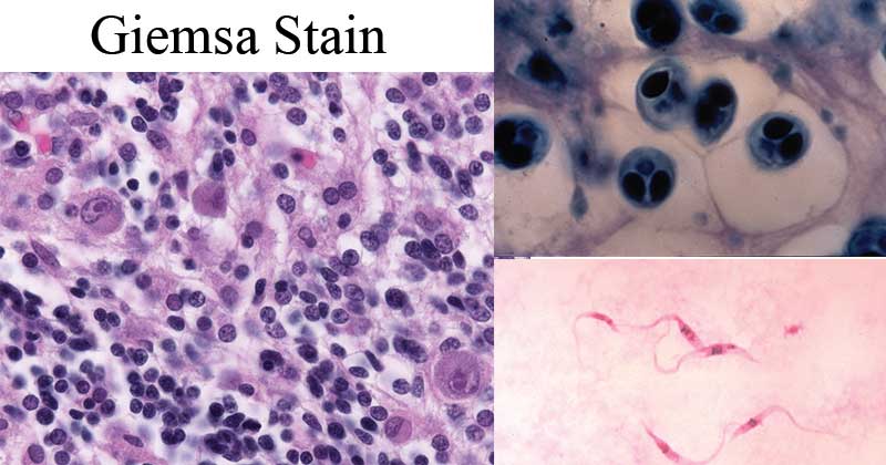
- Diagnosis of Malaria: Giemsa stain is one of the most common diagnostic tools for malaria, a parasitic disease transmitted by mosquitoes. Giemsa stain is used to stain thin and thick blood smears to identify the presence of the Plasmodium parasite within red blood cells.
- Hematology: Giemsa stain is an essential tool in the field of hematology for the identification and differentiation of various types of blood cells. This includes the identification of red blood cells, white blood cells, and platelets, as well as the detection of abnormalities in blood cells, such as sickle cell anemia and leukemia.
- Microbiology: Giemsa stain is used to visualize bacteria, fungi, and other microorganisms, allowing for their identification and analysis. For example, Giemsa stain is used in the diagnosis of diseases caused by bacteria such as Borrelia burgdorferi (the bacteria that causes Lyme disease), and Chlamydia trachomatis (the bacteria that causes sexually transmitted infections).
- Chromosome Analysis: Giemsa stain is used in chromosome analysis to visualize the structure and behavior of chromosomes during cell division. The stain is used to prepare karyotypes (ordered sets of chromosomes) and to detect chromosomal abnormalities, such as aneuploidy, translocations, and inversions.
- Parasitology: Giemsa stain is used in the field of parasitology to visualize parasites and their various stages of development. This includes the detection and diagnosis of parasitic infections caused by protozoa (e.g. Plasmodium, Trypanosoma, and Leishmania) and helminths (e.g. Schistosoma and Ascaris).
- Veterinary Medicine: Giemsa stain is used in veterinary medicine to diagnose various diseases in animals. For example, the stain can be used to diagnose tick-borne diseases, such as ehrlichiosis and babesiosis, in dogs and other animals.
- Plant Biology: Giemsa stain is used in the study of plant cells to visualize the nucleus and other cellular structures. The stain can be used to analyze the chromosomal abnormalities that occur in plants, such as aneuploidy and polyploidy.
Advantages of Giemsa stain:
- Giemsa stain is a simple and easy-to-perform staining method that can be used to prepare and analyze biological samples.
- It can be used for the identification of a wide variety of microorganisms, including bacteria, viruses, and parasites, making it a versatile tool in microbiology and infectious disease research.
- It is a relatively inexpensive staining method, requiring only basic laboratory equipment and reagents.
- Giemsa stain can also be used to visualize the morphology of cells, aiding in the identification of abnormal cells and diagnosis of certain diseases.
- It is a stable staining method, with the stain being long-lasting and resistant to fading.
Disadvantages of Giemsa stain:
- The quality of staining can be affected by variations in staining conditions, including pH, temperature, and staining time, which can lead to inconsistent results.
- Giemsa stain is not specific to particular cell types or structures, making it less effective for some applications compared to other more specific staining methods.
- The staining process can be time-consuming, especially when staining large numbers of samples.
- The stain is not effective for all types of microorganisms, with some species not staining well with Giemsa stain.
- The interpretation of Giemsa-stained samples can be subjective, and inexperienced observers may have difficulty in distinguishing between different cell types or microorganisms.
YOU MAY READ: