Organs of the immune system are the “Thymus, Bone marrow, Spleen and Lymph nodes”
In our Immune system, several morphologically and functionally diverse organs are present and they have various functions in the development of immune responses these lymphoid organs can be distinguished by functions such as the-
A) Primary lymphoid organs – The thymus and bone marrow is the primary or central lymphoid organs where the maturation of lymphocytes takes place.
B) Secondary lymphoid organs – Lymph nodes, spleen, and various mucosal-associated lymphoid tissues (MALT) such as the gut-associated lymphoid tissue (GALT) are the secondary or peripheral lymphoid organs that can trap antigen and provides the sites for mature lymphocytes to interact with that antigen.
Primary lymphoid organs
Once mature lymphocytes have been generated in the primary lymphoid organ (bone marrow), they circulate in the blood and lymphatic system, a network of vessels that collect fluid that has escaped into the tissues from the capillaries of the circulatory system and ultimately return it to the blood.
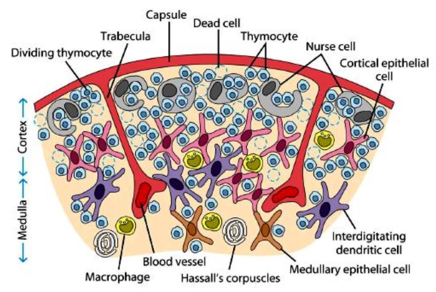
1) Thymus
- Thymus is a flat, bilobed. grayish. a lymphoepithelial organ located just above the heart and beneath the breast bone.
- It is a specialized organ of the immune system Histologically, it is made up of two lobules each lobe of the thymus is divided into a central medulla and a peripheral cortex which is surrounded by an outer capsule.
- These lobules are separated from each other by strands of connective tissue called trabeculae. The cortex and medulla play different roles in the development of T-cells.
- Cells in the thymus can be divided into thymic stromal cells and cells of hematopoietic origin (derived from bone marrow resident hematopoietic stem cells).
- Developing T-cells are referred to as thymocytes and are of hematopoietic origin. Stromal cells include thymic cortical epithelial cells, and thymic medullary epithelial cells. and dendritic cells.
Cortex
- The outer compartment of each lobule is called the cortex and this cortex is densely packed with immature T-cells called thymocytes.
- The cortical portion is mainly composed of lymphoid cells, supported by a network of finely-branched epithelial reticular cells, which is continuous with a similar network in the medullary portion.
- This network forms an adventitia to the blood vessels. The cortex is the location of the earliest events in thymocyte development, where T cell receptor gene rearrangement and positive selection take place.
Medulla
- The inner compartment of each lobule of the thymus is called as the medulla. The medulla is sparsely populated with thymocytes.
- The medulla contains mature, virgin T-cells that have emigrated from the cortex and the layers of comparatively large epithelial cells.
- In the medullary portion, the reticulum is coarser than in the cortex, the lymphoid cells are relatively fewer in number, and there are concentric, nest-like bodies called Hassall’s corpuscles.
- These concentric corpuscles are composed of a central mass, consisting of one or more granular cells, and a capsule formed of epithelioid cells.
- Each follicle is surrounded by a vascular plexus, from which vessels pass into the interior, and radiate from the periphery toward the center, forming a second zone just within the margin of the medullary portion.
- In the center of the medullary portion, there are very few vessels, and they are of minute size. The medulla is the location of the latter events in thymocyte development.
- Thymocytes that reach the medulla have already successfully undergone T cell receptor gene rearrangement and positive selection, and have been exposed to a limited degree of negative selection.
- The medulla is specialized to allow thymocytes to undergo additional rounds of negative selection to remove auto-reactive T-cells from the mature repertoire.
- The gene AIRE is expressed by the thymic medullary epithelium and drives the transcription of organ-specific genes such as insulin to allow maturing thymocytes to be exposed to a more complex set of self-antigens than is present in the cortex.
Functions of thymus
i) Development of CMI – The thymus is a primary lymphoid organ it is the site of T-cell development and maturation.
ii) Maturation of T cells – The function of the thymus is to generate and select a repertoire of T-cells that will protect the body from infection and will recognize antigen- MHC complexes and a small portion react with a combination of self antigen-MHC complexes.
iii) Stock of lymphocytes – The stock of T-lymphocytes is built up in early life, so the function of the thymus is diminished in adults.
iv) Recognition of foreign antigens – The ability of T cells to recognize foreign antigens is mediated by the T cell receptor. The T-cell receptor undergoes genetic rearrangement during thymocyte maturation, resulting in each T-cell bearing a unique T-cell receptor. specific to a limited set of peptide -MHC combinations.
v) Protection from infections – T cells effectively protect the host from various infections. An improperly functioning immune system can cause discomfort, disease, or even death. The type of malfunction falls into one or more of the following major groups: hypersensitivity or allergy, autoimmune disease, or immunodeficiency.
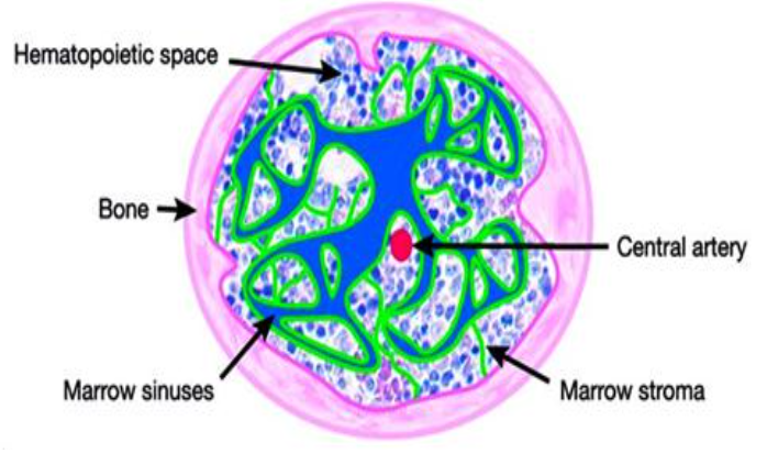
2) Bone Marrow
- Bone marrow is one of the primary lymphoid organs it is the site of B-cell origin and development in humans and mice also.
- Redbone marrow serves as the major source of all blood cells including lymphocytes (in adults). Bone marrow is found in the cavities of most bones in the body including the skull. ribs, sternum, femur and spine.
- Bone marrow is not the site of B-cell development in all species. In birds, a lymphoid organ called the bursa of Fabricius is a lymphoid tissue associated with the gut and is the primary site of B- cell maturation.
- As concern to mammals such as primates and rodents, there is no bursa and no single counterpart to it as a primary lymphoid organ and in cattle and sheep the primary lymphoid tissue hosting the maturation, proliferation, and diversification of B-cells early in gestation is the fetal spleen and later in gestation, this function is assumed by a patch of tissue embedded in the wall of the intestine called the ideal Peyer’s patch and which contains a large number of B-cells.
- However, the distribution of red marrow diminishes with age. The red bone marrow is compartmentalized into hemopoietic and vascular areas and the principle architecture of hemopoietic means blood-forming marrow it consists of sinusoids arranged peripherally around a central vein.
Functions bone marrow
i) Hemopoiesis – Bone marrow is the major site for hemopoiesis all major blood cell types are produced in the bone marrow.
ii) Provides microenvironment – Bone marrow provides the microenvironment for the antigen-independent differentiation of B-cells. Bone marrow also provides an antigen-processing environment.
Secondary lymphoid organs
Examples of the secondary lymphoid organ are the lymph nodes, spleen, adenoids, tonsils, lymphoid patches of the gut, and the appendix. These secondary lymphoid organs are the areas where mature T and B-lymphocytes bind to the antigen and undergo further antigen-dependent differentiation. When T and B lymphocytes bind to the antigen and undergo Ag- dependent differentiation the active immune response begins. These all secondary lymphoid organs can trap antigens.
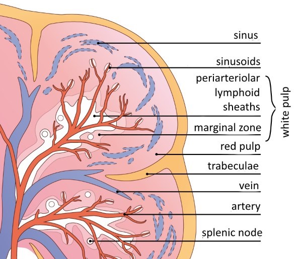
1) Spleen
The spleen is a secondary lymphoid organ. It is a large, ovoid organ situated high on the left of the abdominal cavity. The spleen is surrounded by a capsule that extends several projections (trabeculae) into interior to form a compartmentalized structure. The compartments are of 2 types i.e. red pulp and white pulp. These pulps are separated by a diffused marginal zone.
Red pulp
The spleen consists of a connective tissue network of vascular sinuses called as the red pulp. This red pulp is rich in, macrophages, neutrophils, and erythrocytes (red blood cells), lymphocytes. The red pulp contains few lymphocytes-It is the site where old and defective red blood cells are destroyed and removed. Many of the macrophages within the red pulp contain engulfed red blood cells or iron pigments from degraded hemoglobin.
White pulp
Nestled within the red pulp are compact regions of lymphocytes called as white pulp. B-cells and T-cells occupy separate predetermined areas of the white pulp. The splenic white pulp surrounds the branches of the splenic artery, forming a peri-arteriolar lymphoid sheath (PALS) populated mainly by T-lymphocytes. The primary lymphoid follicles are attached to the periarteriolar lymphoid sheath (PALS). These primary lymphoid follicles are rich in B-cells and some of them contain germinal centers. The marginal zone. located peripheral to the PALS is populated by lymphocytes and macrophages. The initial activation of B-cells and T-cells takes place in the T-cell-rich PALS. Here interdigitating dendritic cells capture antigen and present it to combine with class-second MHC molecules to TH cells. Once activated, B-cells, together with some TH cells, then migrate to primary follicles in the marginal zone.
Functions of spleen
i) Trapping of Ag – The spleen specializes in filtering blood and trapping blood-borne antigens, thus it can respond to systemic infections.
ii) Phagocytosis – The spleen serves as an important station for the phagocytosis of foreign matter and immune reactions against bacteria.
iii) Removal of effete cells – Spleen serves to remove and break down worn (those cells which have completed their life span) erythrocytes.
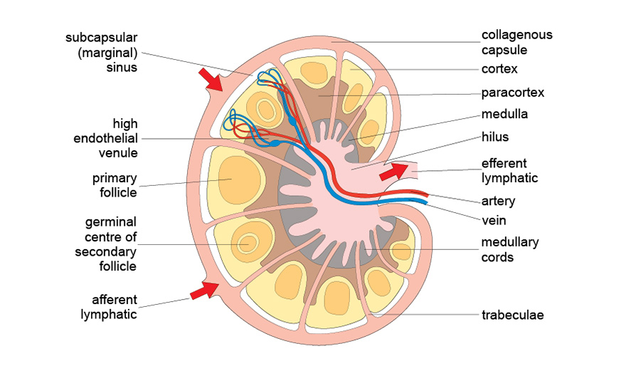
2) Lymph nodes
Lymph nodes are the sites where the immune response is mounted to Ag in the lymph. These are encapsulated bean-shaped structures containing a reticular network packed with lymphocytes, macrophages, and dendritic cells. Lymph nodes are clustered at junctions of the lymphatic vessels.
As lymph percolates through a node, any particulate antigen that is brought in with the lymph will be trapped by the cellular network of phagocytic cells and dendritic cells.
Morphologically. a lymph node can be divided into three regions –
1) The Cortex – The cortex is the outermost layer that contains lymphocytes (mostly B cells), macrophages, and follicular dendritic cells arranged in primary follicles. After the antigenic challenge, the primary follicles enlarge into secondary follicles, each containing a germinal center. In children with B-cell deficiencies, the cortex lacks primary follicles and germinal centers.
2) Paracortex- Beneath the cortex is the paracortex, which is populated largely by T lymphocytes and also contains interdigitating dendritic cells. Lymph nodes taken from neonatally thymectomized mice have unusually few cells in the paracortical region: the paracortex is therefore sometimes referred to as a thymus-dependent area in contrast to the cortex, which is a thymus-independent area.
3) Medulla- The innermost layer of a lymph node, the medulla, is more sparsely populated with lymphoid-lineage cells: of those present, many are plasma cells actively secreting antibody molecules.
 Function of lymph nodes
 Function of lymph nodes
1) Site for immune response – Lymph nodes are the sites where the immune response is mounted to Ag.
2) Filter – Lymph nodes act as filters for filtering out materials that have entered the lymph and providing appropriate cells and niches for immune reactions.
3) Storage of cells – The cortex area of lymph nodes is rich in B cells, Macrophages, and dendritic cells.
4) Ag presentation – The Paracortex area of lymph nodes is rich in T lymphocytes and dendritic cells. These dendritic cells express high levels of class II MHC molecules, which are necessary for presenting antigens to TH cells.
5) Contains Ab-secreting plasma cells – The medulla of lymph nodes is more sparsely populated with plasma cells actively secreting antibody molecules.
References: