Phase contrast microscopy: Definition, principle, parts, uses
A microscope is an optical instrument made of a combination of lenses and used for the magnification of objects. With the advent of high-resolution microscopes, modern microbiologists have access to a microscope. That produces images with high clarity and magnification. The Leeuwenhock’s single-lens microscope has been transformed into a high-resolution multi-lens combination with magnification up to two thousand times.
Phase Contrast Microscope
- This microscope was developed by Fritz Zernikes (1935), a Dutch physicist who was awarded Nobel Prize in 1953 for this contribution. It is a conventional light microscope fitted with a phase-contrast, objective, and a phase-contrast condenser. The phase-contrast microscope is based on the fact that the rate at which light travels through objects is inversely related to their refractive indices.
- Thus, the light passing through one object into another object of a slightly different refractive index undergoes a change in phase. These differences in phase are translated into variation in brightness of the objects and hence the objects differing even slightly in refractive index are viewed by the eye.
- The phase-contrast microscope helps to view living unstained structures of microbial cells. Unlike the interference contrast microscope, the phase-contrast microscope relies upon a single beam of light.
Principle
Highly refractive structures bend light to a much greater angle than do structures of low refractive index. The same properties that cause the light to bend also delay the passage of light by a quarter of a wavelength or so. In a light microscope in bright field mode, light from highly refractive structures bends farther away from the center of the lens than light from less refractive structures and arrives about a quarter of a wavelength out of phase.
Light from most objects passes through the center of the lens as well as to the periphery. Now if the light from an object to the edges of the objective lens is retarded a half wavelength and the light to the center is not retarded at all, then the light rays are out of phase by a half wavelength.
They cancel each other when the objective lens brings the image into focus. A reduction in the brightness of the object is observed. The degree of reduction in brightness depends on the refractive index of the object.
Working
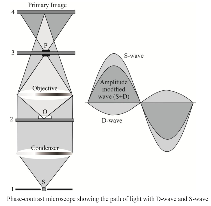
- To get a better understanding of how phase contrast illumination works, we study two wavefronts. This figure simplifies a few things. First, the condenser annulus is just a small aperture located in the center (see the plane labeled ‘1’) and the phase plate is also just covering a small aperture (located in the plane labeled ‘3’). Second, the optical system is greatly simplified by showing only two single lenses to represent all optical elements.
- The plane labeled ‘1’ is the front focal plane of the condenser. The light emanating from the small aperture ‘S’ is captured by the condenser and emerges as light with only parallel wavefronts from the condenser.
- When these plane waves (parallel wavefronts) hit the phase object ‘O’ (located in the object plane labeled ‘2’). some of this light is diffracted (and or refracted) while moving through the specimen.
- Assuming that the specimen does not significantly alter the amplitudes of the incoming wavefronts but mainly changes phase relations with respect to the “unperturbed” wavefronts. newly generated spherical wavefronts that are retarded by 90° (4/4) emanate from ‘O’ (see the purple area that contains now “unperturbed” plane waves and spherical wavefronts).
- It is important to note that there are now two types of waves. the surround wave or S-wave and the diffracted wave or D-wave. which have a relative phase-shift of 90° (1/4). – The objective focuses the D-wave inside the primary image plane (labeled ‘4’), while it focuses the S-wave inside the back focal plane (labeled ‘3’).
- The location of the phase plate ‘P’ has now a profound impact on the S-wave while leaving most of the D-wave “unharmed”. In what is known as positive phase contrast optics, the phase plate ‘P’ reduces the amplitude of all light rays traveling through the phase annulus (mainly S-waves) by 70 to 90% and advances the phase by yet another 90° (A/4).
- However. the phase plate leaves most of the D-waves “untouched”. Hence the recombination of these two waves (D + S) in the primary image plane (labeled ‘4’) results in a significant amplitude change at all locations where there is a now destructive interference due to a 180° (A/2) phase-shifted D-wave.
- The net phase shift of 180° (A/2) results directly from the 90° (4/4) retardation of the D-wave due to the phase object and the 90° (4/4) phase advancement of the S-wave due to the phase plate.
- Without the phase plate. there would be no significant destructive interference that greatly enhances contrast. With phase contrast illumination “invisible” phase variations are hence translated into visible amplitude variations.
- The destructive interference is illustrated in the figure to the right. Blue and orange indicate D-wave and S-wave, respectively. The resulting wave (D + S). has reduced amplitude.
Applications of PCM
1. In various fields – The phase contrast microscope have wide applications in the fields of molecular and cellular biology, microbiology. and medical research.
2. Estimation of intracellular components – Phase contrast microscopy is useful to estimate the concentration of a substance within the cell or cell range.
3. Wet mount and hanging drop – It is useful for examining wet mount of hanging drop preparations for the observation of the internal structure of specimen in living condition.
4. Growth, cell division, and cell movement – It is useful for the examining growth and cell division in bacteria, flagella movement, intestinal and other protozoa such as Amoeba, Trichomonas, etc., living blood cells. cytopathic effects of viruses on tissues.
5. Intracellular components – Useful in the study of bacterial components like endospores, inclusion body containing Poly-beta-hydroxybutyric acid (PHB), polymetaphosphate, and sulfur granules. They are visible because they have a refractive index different from water.
6. Correct cellular structure – A phase-contrast microscope is of the great advance in the study of exact cellular structure. Staining and fixation cause shrinkage and do not reveal the correct structural details. Whereas a phase-contrast microscope gives a sharp outline and correct structure and measurement.
7. Observation of cells in living conditions – One of the major advantages of phase contrast microscopy is that living cells can be examined in their natural state without being killed, fixed, and stained. As a result, the dynamics of ongoing biological processes in live cells can be observed and recorded in high contrast with the sharp clarity of minute specimen detail. Specimens that can be observed and studied include live microorganisms such as protozoa, erythrocytes, bacteria, molds and sperm, thin tissue slices, lithographic patterns, fibers, glass fragments, and sub-cellular particles such as nuclei and organelles.
Advantages of PCM
1. The capacity to observe living cells and, as such, the ability to examine cells in a natural state.
2. Observing a living organism in its natural state and/or environment can provide far more information than specimens that need to be killed, fixed, or stain to view under a microscope.
3. High-contrast, high-resolution images.
4. Ideal for studying and interpreting thin specimens.
5. Ability to combine with other means of observation, such as fluorescence.
6. Modern phase contrast microscopes, with CCD or CMOS computer devices, can capture photo and/or video images.
In addition, advances to the phase-contrast microscope, especially those that incorporate technology, enable a scientist to hone in on the minute internal structures of a particle and can even detect a mere small number of protein molecules.
Disadvantages
1. Annuli or rings limit the aperture to some extent, which decreases the resolution
2. This method of observation is not ideal for thick organisms or particles
3. Thick specimens can appear distorted
4. Images may appear grey or green, if white or green lights are used, respectively, resulting in the poor photomicrograph
5. Shade-off and halo effect referred to phase artifacts