Introduction
Bacterial cells are encased by a multilayered envelope that provides shape, protection, and selective permeability. The innermost barrier is the cytoplasmic (plasma) membrane, a phospholipid–protein bilayer that mediates nutrient transport and energy generation. Surrounding this, most bacteria possess a peptidoglycan cell wall of varying thickness and composition—thick in Gram-positive, thin plus an outer membrane in Gram-negative, and unique mycolic-acid–rich walls in acid-fast species. In some taxa (e.g., mycoplasmas), the peptidoglycan layer is absent. Together, these structures define the cell envelope, with specialized polymers (teichoic acids, lipopolysaccharides) and compartments (periplasm) that confer rigidity, charge, and interactive functions (e.g., virulence, biofilm formation).
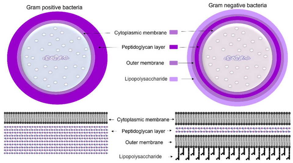
Cell Wall Structure
Gram-Positive Cell Wall
- Peptidoglycan thickness: Very thick (20–80 nm), multiple stacked glycan–peptide layers.
- Teichoic and lipoteichoic acids: Polyalcohol phosphate polymers interwoven in the peptidoglycan; lipoteichoic acids anchor to the membrane, imparting negative charge and cation binding.
- Surface layers (S-layers) and capsules: Proteinaceous S-layers and polysaccharide capsules often coat the peptidoglycan, aiding in adhesion and immune evasion.
Gram-Negative Cell Wall
- Peptidoglycan thickness: Thin (2–7 nm), single or few glycan–peptide sheets in the periplasmic space.
- Outer membrane: Asymmetric bilayer—inner leaflet of phospholipids; outer leaflet rich in lipopolysaccharide (LPS), which defines antigenic specificity and provides a strong permeability barrier.
- Porins and transporters: Trimeric β-barrel porins permit diffusion of small solutes; specific transport proteins mediate uptake of nutrients.
- Periplasm: Gel-like compartment containing peptidoglycan and enzymes (e.g., β-lactamases, binding proteins) that preprocess substrates.
Acid-Fast Cell Envelope (Mycobacteria)
- Mycolic acids: Very long-chain fatty acids covalently linked to arabinogalactan–peptidoglycan complex, forming a waxy barrier.
- Pseudo-periplasm: A poorly defined compartment between the cytoplasmic membrane and the mycolic-acid layer.
- Ziehl–Neelsen staining: Retention of red dye due to mycolic acids confers acid-fastness.
Wall-Less Bacteria
- Mollicutes (e.g., Mycoplasma, Ureaplasma): Lack peptidoglycan; rely on sterol-stabilized plasma membranes for integrity.
- L-forms: Derived from walled bacteria under stress; exhibit fragile, pleiomorphic shapes.
Membrane Structure and Composition
Cytoplasmic (Plasma) Membrane
- Phospholipid bilayer: Roughly equal protein and lipid content; fatty acyl chains vary in length and saturation to modulate fluidity.
- Integral and peripheral proteins: Transporters, receptors, energy-conserving complexes (e.g., ATP synthase), signal transducers.
- Membrane domains: Microdomains enriched in cardiolipin or specific proteins for localized functions (e.g., cell division sites).
Outer Membrane of Gram-Negative Bacteria
- LPS structure: Lipid A anchor plus core oligosaccharide and O-antigen repeats—major endotoxin component.
- Barrier function: Impermeable to many antibiotics and hydrophobic molecules; porins allow passive diffusion of small solutes.
- Maintenance systems: Mla, Lpt pathways ensure asymmetry and repair of outer membrane.
Functional Roles
- Structural integrity: Resists internal turgor pressure (up to ∼3 atm) and external stresses.
- Selective permeability: Membranes regulate nutrient uptake, waste export, and signal transduction; cell wall acts as size filter.
- Pathogenic interactions: Surface polymers (LPS, teichoic acids, mycolic acids) interact with host immune systems; capsules and S-layers modulate virulence.
- Antibiotic targets: Peptidoglycan synthesis enzymes (e.g., PBPs), LPS assembly, membrane integrity pathways are key antibiotic targets.
FAQ (Frequently Asked Questions)
Q1: Why is peptidoglycan absent in some bacteria?
A: Certain obligate intracellular or wall-less bacteria (e.g., Chlamydiaceae, Mollicutes) have lost the peptidoglycan layer, relying instead on host-derived or sterol-stabilized membranes for integrity.
Q2: How do Gram-negative bacteria assemble their outer membrane?
A: Lipopolysaccharides and phospholipids are synthesized at the inner membrane and transported across the periplasm via dedicated systems (e.g., Lpt for LPS, Mla for phospholipids).
Q3: What role do teichoic acids play beyond charge?
A: Teichoic acids bind divalent cations (e.g., Mg²⁺), contribute to cell division and autolysin regulation, and serve as receptors for certain bacteriophages.
Q4: How can the cell envelope be visualized?
A: Transmission electron microscopy (TEM) and cryo-EM reveal layered structures; fluorescent probes distinguish peptidoglycan and membrane domains.
Q5: Why are mycobacterial envelopes particularly impermeable?
A: The long-chain mycolic acids and associated glycolipids create a dense, hydrophobic barrier, reducing permeability and contributing to antibiotic resistance.
References and Links
- Cell envelope (Wikipedia): https://en.wikipedia.org/wiki/Cell_envelope
- Bacterial Cell Wall (ScienceDirect Topics): https://www.sciencedirect.com/topics/biochemistry-genetics-and-molecular-biology/bacterial-cell-wall
- Bacterial Cell Mechanics (PMC, PubMed Central): https://www.ncbi.nlm.nih.gov/pmc/articles/PMC6260806/
- Bacteria Cell Membrane Composition (Frontiers in Molecular Biosciences): https://www.frontiersin.org/articles/10.3389/fmolb.2021.634438/full
- The Gram-Positive Bacterial Cell Wall (Microbiology Spectrum, ASM): https://journals.asm.org/doi/10.1128/microbiolspec.gpp3-0044-2018
- Plasma Membrane–Cell Wall Feedback (Journal of Bacteriology, ASM): https://journals.asm.org/doi/10.1128/jb.00433-22
- Bacterial Cell Wall Structure and Dynamics (Frontiers in Microbiology): https://www.frontiersin.org/research-topics/7085/bacterial-cell-wall-structure-and-dynamics
- Bacterial Membranes: Structure, Domains, and Function (Annual Reviews in Microbiology): https://www.annualreviews.org/doi/10.1146/annurev-micro-102215-095630
- Mollicutes: Cell Envelope Adaptations (Microbiology Reviews): https://www.ncbi.nlm.nih.gov/pubmed/12626688
- Mycobacterial Cell Envelope (FEMS Microbiology Reviews): https://academic.oup.com/femsre/article/28/5/645/455050