Cell Wall of Bacteria
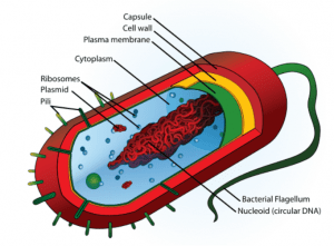
A cell wall is a structure present in plants, fungi, bacteria, algae, and archaea situated outside the cell membrane. Bacteria derive structural support from a peptidoglycan cell wall consisting of disaccharides and amino acids. It is important to remember that there is not a cell wall for all bacteria. However, having said that, it is also important to remember that most bacteria (about 90%) have a cell wall, and they normally have a cell wall. The cell wall of bacteria functions as a support to the cell, and also maintaining the cellular integrity of the cell and protection and any kind of mechanical stress. They are of two types: gram-positive cell wall or gram-negative cell wall.
OVERVIEW OF BACTERIAL CELL WALLS
- A cell wall, not just of bacteria but for all organisms, is found outside of the cell membrane.
- It’s an extra layer that, by providing a semi-rigid form, usually gives the strength that the cell membrane lacks.
- An element known as peptidoglycan (also known as murein) is found in both gram-positive bacteria and gram-negative bacteria cell walls.
- Archaea may be made up of protein, polysaccharides, or molecules similar to peptidoglycan, but they never contain murein. The Bacteria are distinguished from the Archaea by this feature.
- No other location on Earth, other than the cell walls of bacteria, has encountered this specific material.
- But all of these bacterial cell walls are types often contain extra components, rendering the bacterial cell wall a complex structure overall, particularly as compared to eukaryotic microbes’ cell walls.
- Usually, the cell walls of eukaryotic bacteria consist of one single element, such as cellulose in algal cell walls or chitin in fungal cell walls.
- In addition to supplying the cell with overall strength, the bacterial cell wall serves other functions as well. It also helps to preserve the structure of the cell, which is important for how the cell expands, reproduces, gains nutrients, and goes.
- When the cell travels from one setting to another or transports nutrients from its surroundings, it prevents the cell from osmotic lysis.
- Cell walls of bacteria lack a membrane bound nucleus.

The cell wall of bacteria contains peptidoglycan since it is an ingredient that both bacterial cell walls have in common.
STRUCTURE OF PEPTIDOGLYCAN
Peptidoglycan is present outside the cytoplasmic membrane is an integral and basic part of the envelope of the bacterial cell which forms a mesh-like sheet.
- The peptidoglycan network consists of two non-equivalent chains being assembled in two perpendicular directions.
- It gives the cell mechanical rigidity, strengthens the cytoplasmic membrane, and defines the structure of the cells and It acts as a scaffold to anchor other elements of the cell envelope, such as proteins and teichoic acids,
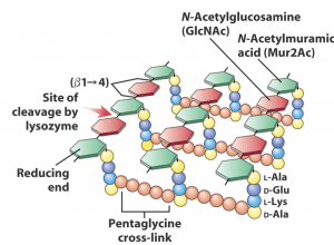
- Peptidoglycan is a polysaccharide made from two long-chain alternative glucose derivatives, N-acetylglucosamine (NAG), and N-acetylmuramic acid (NAM) connected by a beta 1,4-glycoside bond.
- A tetrapeptide that stretches off the NAM sugar unit cross-links the chains to each other, causing a lattice-like structure to form.
- L-alanine, D-glutamine, L-lysine, or meso-diaminopimelic acid (DPA), and D-alanine are the four amino acids that make up the tetrapeptide.
- The D-lactoyl group of each MurNAc residue is substituted by a peptide stem whose composition is most often L-Ala-γ-D-Glu-meso-A2pm (or L-Lys)-D-Ala-D-Ala (A2pm, 2,6-diaminopimelic acid) in nascent peptidoglycan, the last D-Ala residue being lost in the mature macromolecule.
- In the bacterium’s cytosol, the peptidoglycan monomers are synthesized where they bind to a membrane carrier molecule called bactoprenol.
- Bactoprenols transport the monomers of peptidoglycan through the cytoplasmic membrane and work with the enzymes to inject the monomers into existing peptidoglycan, allowing the growth of bacteria after binary fission.
- Glycosidic bonds then connect these monomers into the rising peptidoglycan chains until the new peptidoglycan monomers are added. By way of peptide cross-links between the peptides coming from the NAMs, these long sugar chains are then bound to each other.
- A group of periplasmic enzymes, which are transglycosylases, transpeptidases, and carboxypeptidases, mediate the assembly of peptidoglycan on the outside of the plasma membrane. During the assembly of the murein cell wall, the mechanism of action of penicillin and associated beta-lactam antibiotics is to block transpeptidase and carboxypeptidase enzymes. Therefore, in bacteria, the beta-lactam antibiotics are said to “block cell wall synthesis.”
- The hydrolysis of β1 → 4-linkages between N-acetylmuramic acid and N-acetyl-β-glucosamine residues in peptidoglycan is catalyzed by Lysozyme, also called muramidase (peptidoglycan N-acetylmuramoyl hydrolase).

Cell wall of bacteria gram-positive and negative
GRAM POSITIVE CELL WALLS
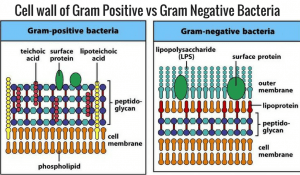
- The cell wall of bacteria of gram-positive is thick (15-80 nanometers), consisting of several layers of peptidoglycan.
- Peptidoglycan is primarily composed of the cell walls of gram-positive bacteria.
- Gram-positive cell walls retain purple crystal violet dye when subjected to the Gram-staining procedure.
- Up to 90 percent of the cell wall will potentially be peptidoglycan, with layer after layer forming around the cell membrane.
- There is a group of molecules called teichoic acids and lipoteichoic acids unique to the cell wall of Gram-positive.
- Teichoic acids are somehow involved in the outside of the plasma membrane in the regulation and assembly of muramic acid subunits. There are instances in which teichoic acids have been involved in bacterial adherence to tissue surfaces, particularly in streptococci.
- Usually, the NAM tetrapeptide is cross-linked with a peptide underbridge, and it is normal to have complete cross-linking. All this works to create an extremely high wall of cells.
- Many surface proteins (e.g. protein A, fibrinonectin-binding proteins, collagen adhesin) anchored to peptidoglycan and implicated in pathogenic processes are found in Gram-positive bacteria. A membrane protein called sortase A catalyzes the anchoring reaction
GRAM NEGATIVE CELL WALLS
- The cell wall of bacteria of gram-negative is relatively thin (10 nanometers) and is composed of a single layer of peptidoglycan surrounded by an outer membrane.
- The cell walls of gram-negative bacteria, with more ingredients total, are more complex than those of gram-positive bacteria.
- Gram-negative cell walls do not retain purple crystal violet dye when subjected to the Gram-staining procedure
- They also contain peptidoglycan, but they only contain a few layers, about 5-10% of the overall cell wall.
- The presence of a plasma membrane situated outside the peptidoglycan layers, known as the outer membrane, is what is most noteworthy about the gram-negative cell wall. The outer membrane is composed of a lipid bilayer.
- Gram-negative bacteria’s outer membrane invariably contains a unique component, lipopolysaccharide ( LPS or endotoxin), which is poisonous to animals. The outer membrane is generally considered to be part of the cell wall in Gram-negative bacteria.
- Also, the outer membrane creates resistance to many antibiotics.

References
https://bio.libretexts.org/Bookshelves/Microbiology/Book%3A_Microbiology_(Kaiser)/Unit_1%3A_Introduction_to_Microbiology_and_Prokaryotic_Cell_Anatomy/2%3A_The_Prokaryotic_Cell_-_Bacteria/2.3%3A_The_Peptidoglycan_Cell_Wall#:~:text=Peptidoglycan%2C%20also%20called%20murein%2C%20is,of%20the%20NAM%20(Figure%202.3.
http://www.textbookofbacteriology.net/structure_5.htmlhttps://www.creative-proteomics.com/services/peptidoglycan-structure-analysis.htmhttps://academic.oup.com/femsre/article/32/2/149/2683904https://www.sciencedirect.com/topics/chemistry/peptidoglycanhttps://www.sciencedirect.com/topics/agricultural-and-biological-sciences/lysozyme#:~:text=Lysozyme%2C%20also%20called%20muramidase%20(peptidoglycan,constituent%20of%20bacterial%20cell%20walls.
