Introduction
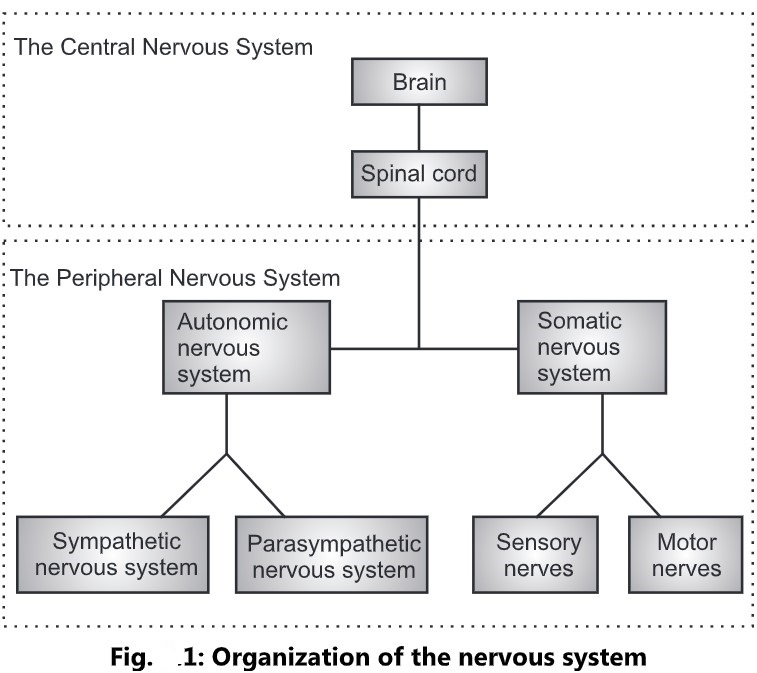
- The two principle divisions of the nervous system are the central nervous system (CNS) and peripheral nervous system (PNS).
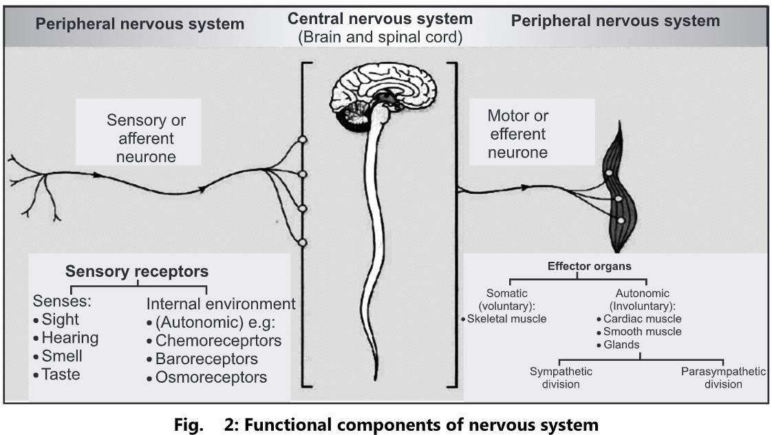
- The CNS consists of brain and spinal cord, integrates and correlates many different kinds of incoming sensory information.
- The CNS is also the source of thoughts, emotions and memories.
- The components of peripheral nervous system (PNS) are subdivided into a somatic nervous system (SNS) and autonomic nervous system (ANS).
- The SNS is voluntary.
- The SNS consists of sensory neurons and motor neurons.
- Sensory neurons: They convey information from somatic receptors in the head, body wall, limbs and from receptors of special senses of vision, hearing, taste and smell to the CNS.
- Motor neurons: They conduct impulses from the CNS to skeletal muscles.
- The ANS is involuntary.
- The ANS consists of sympathetic and parasympathetic division.
- Sensory neuron: They convey information from autonomic sensory receptors, located primarily in the visceral organs such as stomach and lungs to the CNS.
- Motor neuron: They conduct nerve impulses from the CNS to smooth muscles, cardiac muscles and glands.
Autonomic Nervous System
- The peripheral nervous system consists of somatic nervous system (SNS) and autonomic nervous system (ANS).
- Somatic nervous system consists of sensory neurons and motor neurons.
- Sensory neurons convey message from periphery to the CNS.
- These sensations include sensations of pain, temperature, taste, smell, hearing, vision, etc.
- Motor neurons innervate the skeletal muscles and lead to voluntary movements.
- The autonomic or involuntary part of the nervous system which controls the autonomic function of the body.
- It consists of two types of neurons:
- Autonomic sensory neuron
- Autonomic motor neuron
Autonomic Sensory Neuron (Afferent):
- These neurons are associated with interoceptors which are sensory receptors located in blood vessels, visceral organs, and muscles.
- Sensory neurons are responsible for receiving information from sensory receptors to the central nervous system.
Autonomic Motor Neuron (Efferent):
- These regulates visceral activities by either increases or decreases in ongoing activities in their effector tissues (cardiac muscle, smooth muscles or glands). E.g. Change in diameter of pupil, dilation or constriction of blood vessels, adjusting the rate and force of heart rate.
Divisions of Autonomic Nervous System:
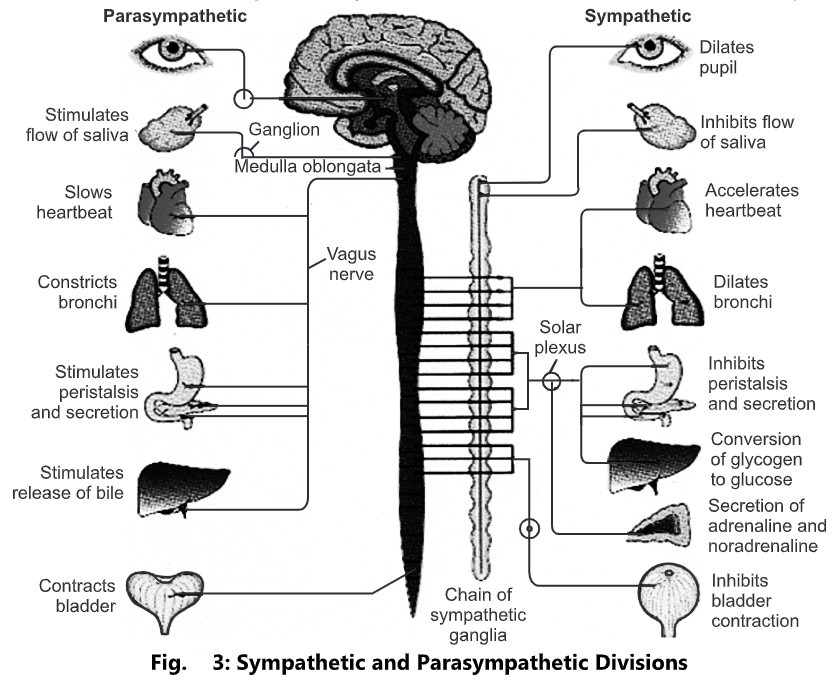
- The autonomic nervous system is separated into two divisions:
- Sympathetic (Thoracolumbar outflow) division
- Parasympathetic (Craniosacral outflow) division
- These two divisions have both structural and functional differences.
- They normally work in the opposite manner.
- Each division has two motor neurons, autonomic ganglia and effector organs.
- These are;
- Pre-ganglionic neurons: The first motor neurons which lies before the ganglion is called as pre-ganglionic neuron. The myelinated axon is called as pre-ganglionic fibre.
- Post-ganglionic neurons: The second motor neuron which lies after the ganglion and terminates in the effector organ is called as post-ganglionic neuron. Its axon is called as post-ganglionic fibres.
- The autonomic ganglion is the collection of cell bodies outside the CNS.
Sympathetic Division
- It is called as thoracolumbar division.
- It consists of two types of autonomic ganglia.
- Sympathetic trunk ganglia
- Prevertebral ganglia
Sympathetic Trunk Ganglia:
- These are the ganglia lie in a vertical row on either side of the vertebral column.
- These lies close to the spinal cord and therefore the pre-ganglionic fibres are short.
Prevertebral Ganglia:
- There are three types of ganglion:
- Coeliac ganglion
- Superior mesenteric ganglion
- Inferior mesenteric ganglion
- These are the ganglion situated close to the abdominal cavity.
- Most of the ganglia of sympathetic trunk are arranged on both sides of the spinal cord.
- Ganglia are close to the CNS and distant from effector organs.
- The pre-ganglionic nerve fibres are shorter.
- The post-ganglionic nerve fibres are longer.
- The pre-ganglionic neurotransmitter is acetylcholine.
- Most of the post-ganglionic nerve fibres are adrenergic.
- The targeted receptors are mostly adrenergic.
- These are distributed throughout the body.
Parasympathetic Division
- It is called as cranio-sacral division.
- It contains parasympathetic ganglia.
- The parasympathetic ganglia are dispersed.
- The ganglia are near or within the wall of the visceral effectors.
- The pre-ganglionic nerve fibres are longer.
- The post-ganglionic nerve fibres are shorter.
- The pre-ganglionic nerve fibres are acetylcholine.
- Most of the postganglionic nerve fibres are cholinergic.
- The targeted receptors are mostly cholinergic.
- The distribution is limited, particularly to heart, viscera of thorax, abdomen and pelvis.
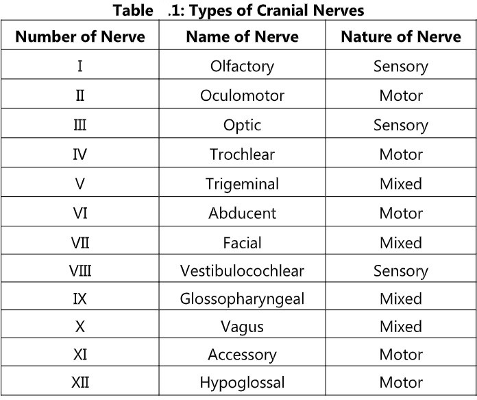
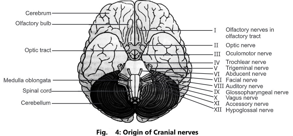
Cranial Nerves
- There are 12 pairs of cranial nerves originating from the nuclei in the inferior surface of the brain.
- Some are sensory, some are motor and some are mixed.
- Their names and numbers are as follows:
Olfactory Nerves:
- It is sensory type of nerve with afferent fibre.
- It originates in the olfactory lobe i.e. root of nose and terminates in the temporal lobe of cerebrum.
- It is associated with sense of smell.
Optic Nerves:
- It is sensory type of nerve with afferent fibre.
- It originates in the retina of eyes and terminates in the vision area of occipital lobe of the cerebrum.
- It is related with sense of vision.
Oculomotor Nerves:
- It is mixed type of nerve with efferent as well as afferent fibre, but primarily it is motor originates in the mid-brain.
- Efferent (motor) portion: It innervates the skeletal muscles it moves the eyeball and innervates the smooth muscles that constrict pupil and lens shape for far and near vision.
- Afferent (sensory) portion: It is related to movement of eyeball and regulating the size of pupil.
Trochlear Nerves:
- It is mixed type of nerve but primarily motor and originates in the midbrain.
- It is the smallest of the 12 cranial nerves.
- The motor portion is related to the movement of eyeball and sensory vision carries information from muscles of eye to midbrain.
Trigeminal Nerves:
- It is a mixed type of nerve fibre.
- It is the largest among all the cranial nerves.
- The motor portion originates from pons and innervates the muscles of mastication (skeletal chewing muscles).
- The sensory portion consists of three branches:
- Ophthalmic nerve: It contains axons from skin of eyelids, eyeball, lacrimal glands, nasal cavity, nose and forehead.
- Maxillary nerve: It contains axons from the mucosa of nose, parts of pharynx, upper teeth, upper lip and lower eyelid.
- Mandibular nerve: It contains axon from tongue, lower teeth, skin over mandible, and cheek.
- Motor function: Chewing
- Sensory function: Conveys impulses for touch, pain and temperature.
Abducens Nerve:
- A mixed type of nerve, but 1° motor that originates in the pons.
- The motor portion innervates the skeletal muscles that move eyeball.
- The sensory portion transmits information from proprioceptors in muscles. It is related to the movement of eyeball and muscles sense (proprioception).
- Motor function: Movement of eyeball
- Sensory function: Proprioception
Facial Nerve:
- It is a mixed type of nerve.
- The motor fibre originates from pons and innervates the skeletal muscle of face, nose, palate, lacrimal and salivary gland.
- The sensory fibre transmits information from taste buds in the tongue and mouth.
- Motor function: Facial expression
- Sensory function: Proprioception and taste
Vestibulocochlear Nerve:
- It is sensory type of nerve that transmits information from receptor in ear.
- It consists of two nerves:
- Vestibular nerve: It arises from semicircular canals of the inner ear and conveys impulses to the cerebellum. They are associated with maintenance of posture and balance.
- Cochlear nerve: It originates in the spiral organ of the inner ear and conveys impulses to the hearing area of cerebral cortex. Cochlear nerve is responsible for hearing.
Glossopharyngeal Nerve:
- It is a mixed type of nerve.
- The motor fibres originate from medulla oblongata and innervate the tongue and pharynx.
- The sensory fibres originate from salivary glands and terminate in the medulla oblongata.
- Motor function: Elevates the pharynx during swallowing and speech.
- Sensory function: Taste, touch, pain and temperature sensations, monitoring of blood pressure.
Vagus Nerve:
- It is a motor type of nerve.
- The motor fibres originates in the medulla oblongata and innervates the smooth muscles of pharynx, larynx, trachea, heart, oesophagus, stomach, intestine, pancreas, gall bladder, bile duct, spleen, kidney, ureter, blood vessels in thoracic and abdominal cavities.
- The sensory fibres convey impulses from same organs to brain.
- Motor function: Swallowing, coughing and voice production
- Sensory function: Taste, touch, pain and temperature sensations, monitoring of blood pressure.
Accessory Nerve:
- It is a mixed type of nerve, primarily motor nerve.
- It originates from the medulla oblongata and innervates the voluntary muscles of pharynx and skeletal muscle of neck.
- Motor function: Neck controls swallowing movements and movement of head and shoulders.
Hypoglossal Nerve:
- It is a mixed type of nerve but primarily motor nerve.
- It originates in the medulla oblongata and supplies to the muscle of tongue.
- The sensory function is gives sensation to tongue.
- Sensory function: Proprioception
- Motor function: Movement of tongue during speech and swallowing.
Spinal Nerves
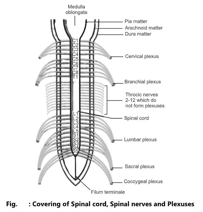
- There are 31 pairs of spinal nerves that leaves the vertebral canal by passing through the intervertebral foramina formed by adjacent vertebrae.
- They are named and grouped according to the vertebrae with which they are associated.
- 8 Cervical
- 12 Thoracic
- 5 Lumbar
- 5 Sacral
- 1 Coccygeal
- Even though there are only seven cervical vertebrae, eight cervical nerves are present because the first pair leaves the vertebral canal between the occipital bone and the atlas and the eighth pair leaves beneath the last cervical vertebra.
- The lumbar, sacral and coccygeal nerves leave the spinal cord near its termination at the level of first lumbar vertebra and extend downwards inside the vertebral canal in the subarachnoid space, forming horse’s tail like structure called as Cauda equina.
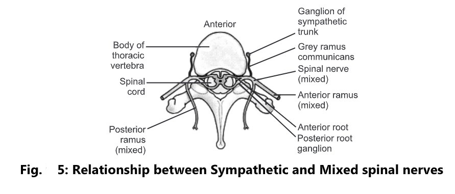
- Atypical spinal nerve has two connections to the cord: Posterior root and Anterior root.
Anterior Nerve Root:
- It consists of motor nerve fibres which are the axons of nerve cells in the anterior column of grey matter in the spinal cord and in the thoracic and lumbar regions, sympathetic nerve fibres which are the axons of cells in the lateral columns of grey matter.
Posterior Nerve Root:
- It consists of sensory nerve fibres.
- Just outside the spinal cord there is a spinal ganglion (posterior root ganglion), consisting of a little cluster of cell bodies.
- Sensory nerve fibres pass through these ganglia before entering the spinal cord.
- The posterior and anterior roots unite to form a spinal nerve at the intervertebral foramen.
- Because the posterior root contains sensory axons and the anterior root contains motor axons, as spinal nerve is classified as a mixed nerve.
- The posterior root contains a posterior root ganglion in which cell bodies of sensory neurons are located.
- After leaving the spinal cord the nerve roots have a covering of dura and arachnoid maters.
- These terminate before the two roots join to form the mixed spinal nerve.
- The nerve roots do not have the pia mater covering.
- After emerging from the intervertebral foramen each spinal nerve divides into a ramus communicans, a posterior ramus and an anterior ramus.
- The rami communicans are part of preganglionic sympathetic neurons of the autonomic nervous system.
- The posterior rami pass backwards and divide into medial and lateral branches to supply skin and muscles of relatively small areas of the posterior aspect of the head, neck and trunk.
- The anterior rami supply the anterior and lateral aspects of the neck, trunk, the upper and lower limbs.
- In the cervical, lumbar and sacral regions the anterior rami unite near their origins to form plexuses (large masses of nerves), where nerve fibres are regrouped and rearranged before proceeding to supply skin, bones, muscles and joints of a particular area.
- There are five large plexuses of mixed nerves formed on each side of the vertebral column.
- They are as follows;
- Cervical plexuses: It is formed by the anterior rami of the first four cervical nerves.
- Brachial plexuses: The anterior rami of the lower four cervical nerves and a large part of the first thoracic nerve form the brachial plexus.
- Lumbar plexuses: It is formed by the anterior rami of the first three and part of the fourth lumbar nerves.
- Sacral plexuses: It is formed by the anterior rami of the lumbosacral trunk and the first, second and third sacral nerves.
- Coccygeal plexuses: The coccygeal plexus is a very small plexus formed by part of the fourth, fifth sacral and the coccygeal nerves.