Physiology of Vision
- The rods and cones are two types of photoreceptors that help in transducing light rays into the receptor potential.
- The retina of each eye contains about 6 millions of cones and 120 millions of rods.
- Rods: These helps in viewing the grey shades in dim light and are responsible for absorbing shapes and movements.
- Cones: They provide colour vision in the bright light. The cones cannot functions in dim light.
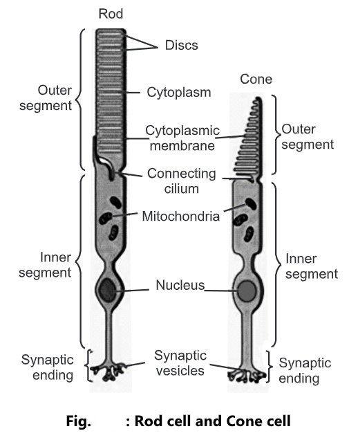
- The rods and cones are named because of their shapes.
- The rods and cones are differentiated into two parts:
- Outer segment
- Inner segment
- The outer segments of cones are cone shaped whereas those of rods are rod shaped.
- The outer segment of rods as well as cones contains invaginated membranes called as discs.
- The first step in visual transduction in absorption of light by a photo pigments. These pigments are coloured protein that undergoes structural changes when they absorbs light, in the outer segment of photoreceptor.
- The pigment present in the rods is rhodopsin and the cones is iodopsin.
- Both rhodopsin as well as iodopsin contains retinal (derivatives of vitamin-A).
- All photo pigments associated with vision contains two parts:
- A glycoprotein known as opsin.
- Derivative of vitamin – A known as retinal.
- Good vision depends on adequate dietary intake of carotene-rich vegetables such as carrot, spinach, broccoli and yellow squash or foods that contains vitamin-A such as liver.
- Retinal is light absorbing part of all visual photopigments.
- In human retina, there are four different opsins, three in the cones and one in the rods (rhodopsin).
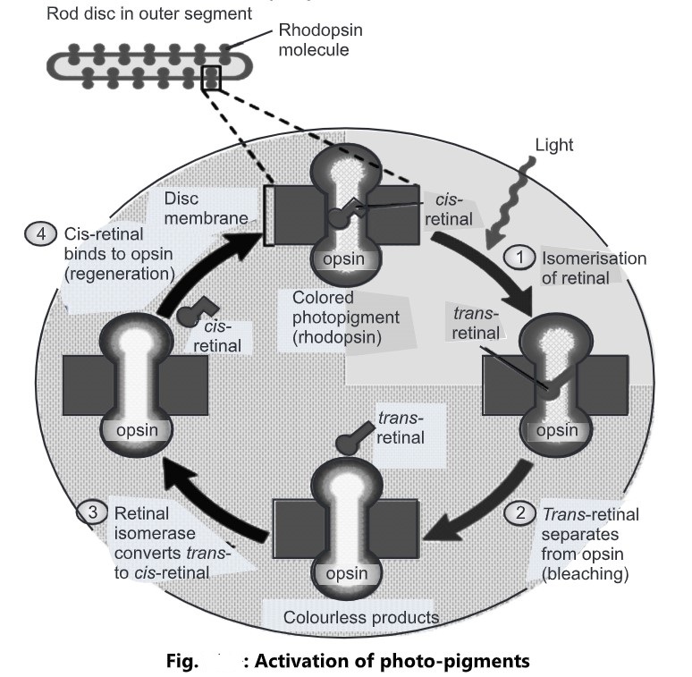
Activation of Photo-pigments:
- Photo-pigments respond to light in the following process:
(1) In the darkness, retinal has a bent shape, called as cis-retinal, which fits tightly into the opsin portion of the photopigment when cis-retinal absorbs a photon of light; it straightens out to a shape called as trans-retinal.
(2) This cis-to-trans conversion is called as isomerisation and is the first step in visual transduction.
(3) An enzyme called as retinal isomerase converts trans-retinal back to cis-retinal.
(4) The cis-retinal then binds to opsin, reforming a functional photopigment. This process of resynthesis of photopigment is called as regeneration.
- The inner segment of rods and cones contains mitochondria, which provide energy.
- The discs of the rods are constantly regenerated whereas this does not occur in cones.
Light and Dark Adaptation:
- When the person emerges from a dark surroundings (a tunnel) into the sunshine, the light adaptation occurs.
- Our visual system adjusts in seconds to a brighter environment by decreasing its sensitivity.
- When the person enters a darkened room such as a theatre, visual system undergoes dark adaptation by increasing the sensitivity.
- As the light level increases more and more photopigment is bleached.
- While light is bleaching, more photopigment molecules, however, others are being regenerated.
Release of Neurotransmitter by Photoreceptors:
Operation of Rod Photoreceptor in Darkness:
- In darkness, the cyclic GMP levels of photoreceptors are high.
- This level of GMP opens the ligand-gated sodium ions channels.
- Inflow of Na+ ions through the channel in the photoreceptor takes place called as dark current which partially depolarizes the photoreceptor.
- As a result, in darkness the membrane potential of a photoreceptor is -30 mV which is much closer to -70 mV.
- This partial depolarization during darkness triggers the release of neurotransmitters at the synaptic terminals.
- The neurotransmitter is the amino acid glutamate or glutamic acid.
- This glutamate inhibits the bipolar cells that synapse with rods.
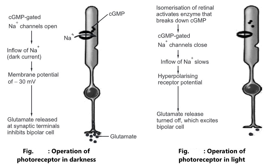
Operation of Rod Photoreceptor in Light:
- When light strikes on the retina the cis-retinal undergoes isomerization to trans-retinal, enzyme transduction get activated which activates phosphodiesterase which breaks down the cyclic GMP and leads to decrease in cyclic GMP levels which in turn is responsible for closing of sodium channels.
- So, inflows of Na+ ions in the photoreceptor get decreases.
- The membrane potential becomes more negative -70 mV (hyperpolarizing receptor potential).
- This sequence of events produces a hyperpolarizing receptor potential that decreases the release of neurotransmitter glutamate.
- Thus, light excites bipolar cells that synapse with rods by turning off the release of inhibition of neurotransmitter.
- The excited bipolar cells subsequently stimulate the ganglion cells to form action potentials in their axons.