Arthrology:
- It is the branch of science concerned with the study of anatomy, function, dysfunction, and treatment of joints and articulations.
Joint (Articulation):
- It is a point of contact between two bones, between bone and cartilage, or between bone and teeth.
- A joint is the site at which any two or more bones articulate or come together.
- Some joints have no movement (fibrous), some have only slight movement (cartilaginous) and some are freely movable (synovial).
Classification of Joint
- Joints are classified on the basis of their structure and function.
(I) Structural Classification of Joints:
- Structural classification is based on the materials that hold the joint together and whether or not a cavity is present in the joint.
- Fibrous joints: There is no synovial cavity and the bones are held together by fibrous tissue.
- Cartilaginous joints: There is no synovial cavity and the bones are held together by cartilage.
- Synovial joints: There is a synovial cavity and the bones are held together by the dense irregular connective tissue of an articular capsule and accessory ligaments.
(II) Functional classification of joints:
- Functional classification is based on the degree to which the joint permits movement.
Synarthrosis:
- It is an immovable joint.
- Structurally, it may be a fibrous or cartilaginous joint.
- The articular surfaces are connected by tough fibrous tissue.
- The edges of the bones are devoted to one another as in the sutures of the skull.
Amphiarthrosis:
- It is a slightly movable joint.
- Structurally, it may be a fibrous or cartilaginous joint.
- A pad of cartilage lies between the bone surfaces.
- There is a fibrous capsule to hold the bone and cartilage in place.
- The cartilage of such joints acts as shock absorbers.
Diarthrosis:
- It is a freely movable joint.
- Structurally, it is a synovial joint.
- The movement of these joints is restricted by the shape of the articulating surfaces and ligaments which hold them together.
- These ligaments are of elastic connective tissue.
Based on the structural differences joints are of the following types:
Fibrous Joint or Fixed Joint
- It lacks a synovial cavity.
- The articulating bones are held together by a dense irregular connective tissue.
- It permits little or no movement.
- The three types of fibrous joints are:
- Sutures
- Syndesmoses
- Interosseous membranes
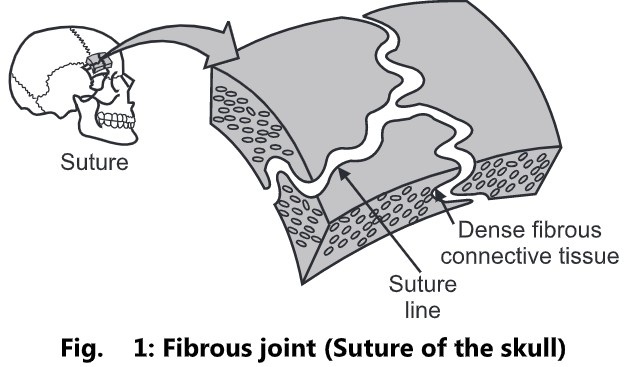
(i) Sutures:
- It is a fibrous joint composed of a thin layer of dense irregular connective tissue.
- It occurs only between the bones of the skull. E.g. Coronal suture between the parietal and frontal bones.
- The irregular and interlocking edges of sutures give them additional strength and decrease the chance of fracturing.
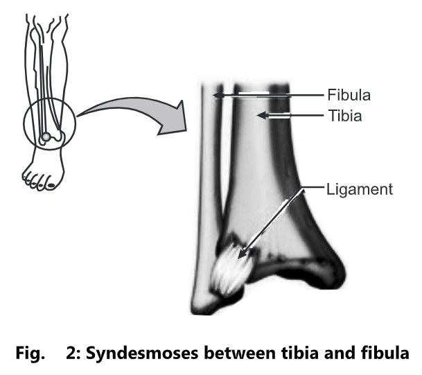
(ii) Syndesmoses:
- In syndesmoses, a greater distance is present between the articulating surfaces.
- It contains denser irregular connective tissue than in a suture.
- The dense irregular connective tissue is typically arranged as a bundle and the joint permits limited movement. E.g. The distal tibiofibular joint.
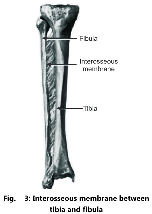
(iii) Interosseous Membranes:
- It contains a sheet of dense irregular connective tissue that binds neighboring long bones.
- It permits slight movement.
- There are two interosseous membrane joints in the human body.
- One occurs between the radius and ulna in the forearm and the other occurs between the tibia and fibula in the leg.
Cartilaginous Joints
The characteristics of cartilaginous joints are:
- It lacks a synovial cavity.
- It allows little or no movement.
- The articulating bones are tightly connected by either hyaline cartilage or fibrocartilage.
- The two types of cartilaginous joints are:
- Synchondroses
- Symphysis
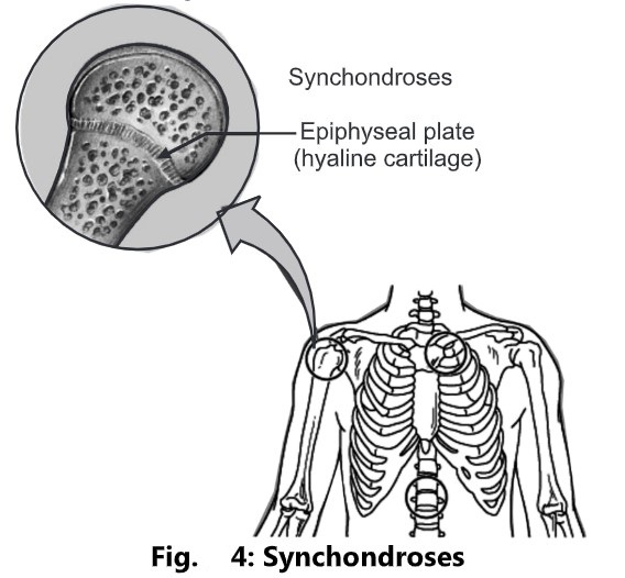
(i) Synchondroses:
- It is a cartilaginous joint in which hyaline cartilage is the connecting material. E.g. The epiphyseal (growth) plate that connects the epiphysis and diaphysis of a growing bone.
- It is a synarthrosis, an immovable joint.
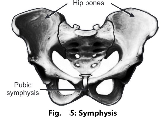
(ii) Symphysis:
- It is a cartilaginous joint in which the ends of the articulating bones are covered with hyaline cartilage, and abroad, a flat disc of fibrocartilage connects the bones.
- All symphysis occur in the midline of the body. E.g. The pubic symphysis between the anterior surfaces of the hip bones.
- It is an amphiarthrosis, a slightly movable joint.
Synovial Joint
- The exclusive characteristic of the synovial joint is the presence of space called a synovial cavity between the articulating bones.
- All synovial joints are classified as diarthrosis or movable joints.
- A synovial joint shows the presence of the following structures:
- Articular or hyaline cartilage
- Capsule or capsular ligament
- Synovial membrane
- Synovial fluid
- Other intracapsular structures
- Extracapsular structures
Articular or Hyaline Cartilage:
- The parts of the bones that are in contact are covered with hyaline cartilage.
- It provides a smooth articular surface that is strong enough to absorb compression forces and bear the weight of the body.
- This leads to increased stress on other structures in the joint.
- It has no blood supply and receives its nourishment from synovial fluid.
Capsule or Capsular Ligament:
- The joint is surrounded by a sheath of fibrous tissue that holds the bones together.
- It is sufficiently loose to allow movement but strong enough to protect from injury.
Synovial Membrane:
- It is composed of epithelial cells.
- This membrane is found in:
- The lining of the capsule
- The parts of bones within the joints not covered by articular cartilage
- All intracapsular structures
Synovial Fluid:
- It is a thick, sticky fluid of egg-white consistency, secreted by synovial membranes into the synovial cavity.
- Functions of synovial fluid:
- It provides nutrients for the structures within the joint cavity.
- It contains phagocytes, which remove microbes and cellular debris.
- It acts as a lubricant.
- It maintains joint stability.
- It prevents the ends of bones from being physically separated.
- The little sacs of synovial fluid are present in some joints. E.g. The knee.
- They act as cushions to prevent friction between a bone and a ligament or tendon, or skin.
Intracapsular Structures:
- Some joints have structures within the capsule, but outside the synovial membrane, that assist in the maintenance of stability. E.g. Fat pads in the knee joint.
- These structures are covered by a synovial membrane.
Extracapsular Structures:
- Ligaments that blend with the capsule provides additional stability at most joints.
- Muscles or their tendons also provide stability and stretch across the joints they move.
Nerve and blood supply:
- Nerves and blood vessels crossing a joint usually supply the capsule and the muscles that move it.
- Synovial joints contain many nerve endings that are distributed to the articular capsule and associated ligaments.
- Arteries and their numerous branches penetrate the ligaments and articular capsule to deliver oxygen and nutrients.
- Veins remove the carbon dioxide and wastes from the joints.
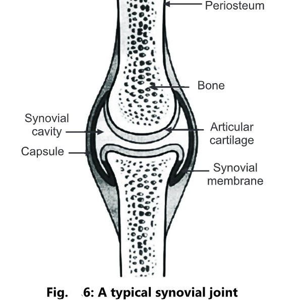
Table 1: Movements of Synovial Joints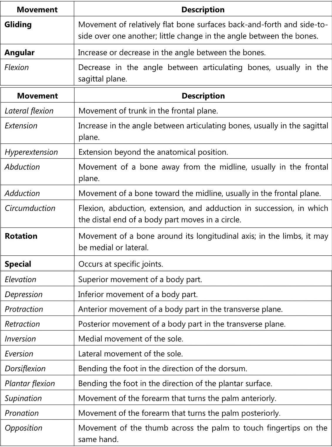
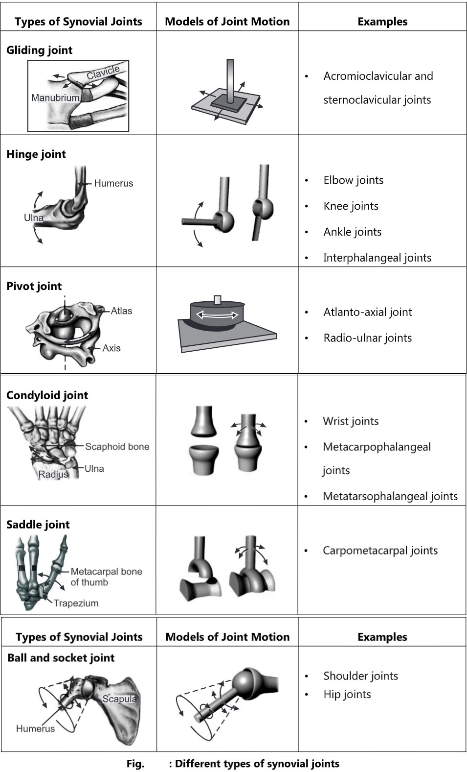
Types of Synovial Joints:
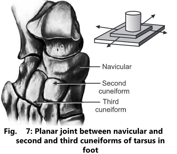
Gliding Joint:
- The articulating surfaces are flat or slightly curved.
- They permit back-and-forth and side-to-side movements between the flat surfaces of bones.
- Many planar joints are biaxial because they permit movement around two axes.
- An axis is a straight line around which a rotating bone moves.
- Examples of planar joints are:
- Intercarpal joints (between carpal bones at the wrist),
- Intertarsal joints (between tarsal bones at the ankle),
- Sternoclavicular joints (between the manubrium of the sternum and the clavicle),
- Acromioclavicular joints (between the acromion of the scapula and the clavicle),
- Sternocostal joints (between the sternum and ends of the costal cartilages at the tips of the second through seventh pairs of ribs),
- Vertebrocostal joints (between the heads and tubercles of ribs and transverse processes of thoracic vertebrae).
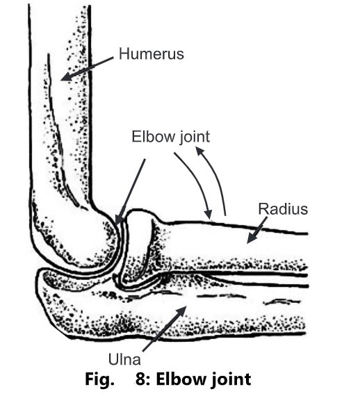
Hinge Joint:
- The convex surface of one bone fits into the concave surface of another bone.
- It produces an angular, opening-and-closing motion.
- In most joint movements, one bone remains in a fixed position while the other moves around an axis.
- These are monaxial because they typically allow motion around a single axis.
- They permit only flexion and extension.
- Examples of hinge joints are:
- Knee joint
- Elbow joint
- Ankle joint
- Interphalangeal joint
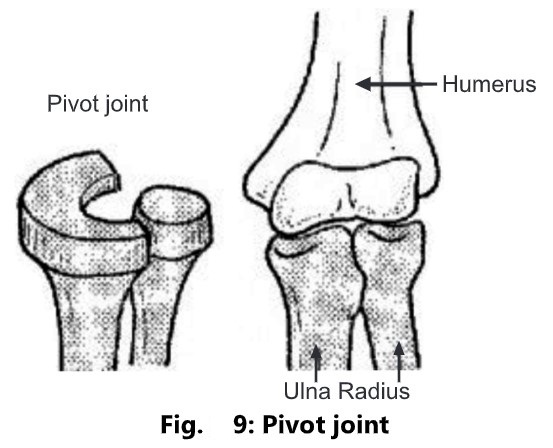
Pivot Joint:
- In a pivot joint, the rounded or pointed surface of one bone articulates with a ring formed by another bone and ligament.
- It is a monaxial joint because it allows rotation only around its own longitudinal axis.
- Examples of pivot joints are;
- Atlanto-axial joint, in which the atlas rotates around the axis and permits the head to turn from side to side.
- Radioulnar joints that enable the palms to turn anteriorly and posteriorly.
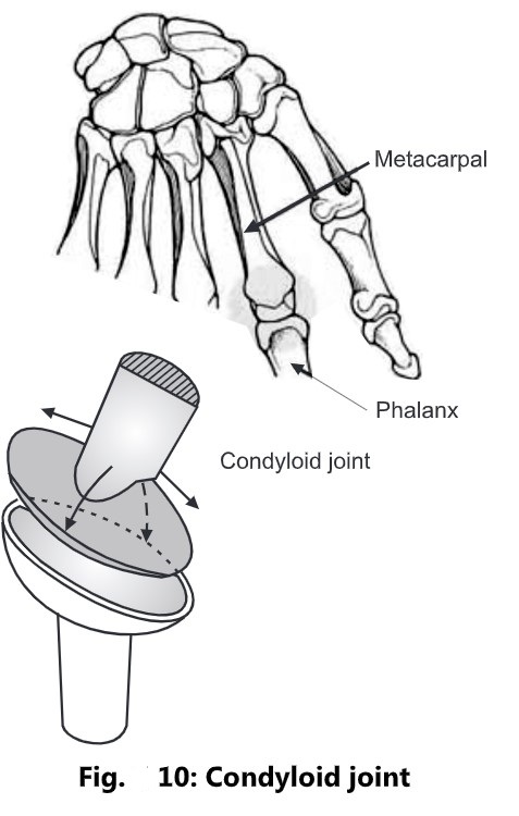
Condyloid Joint:
- In a condyloid joint or ellipsoidal joint, the convex oval-shaped projection of one bone fits into the oval-shaped depression of another bone.
- It is a biaxial joint because the movement it permits is around two axes.
- Examples of condyloid joints are the wrist and metacarpophalangeal joints for the second through fifth digits.
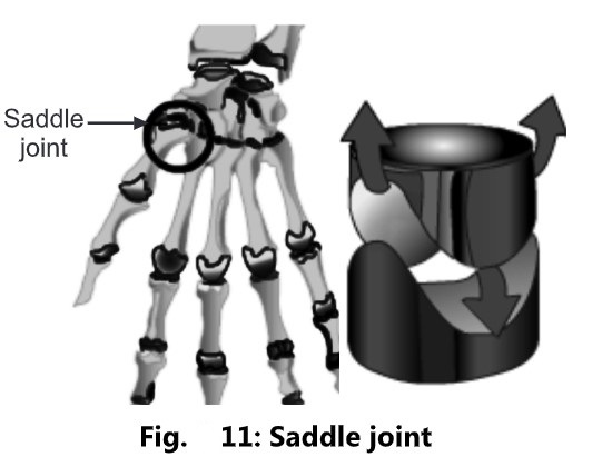
Saddle Joint:
- In a saddle joint, the articular surface of one bone is saddle-shaped, and the articular surface of the other bone fits into the saddle.
- It is a modified condyloid joint.
- These are triaxial joints, permitting the movements around three axes.
- An example of a saddle joint is the carpometacarpal joint between the trapezium of the carpus and the metacarpal of the thumb.
Ball and Socket Joint:
- It consists of a ball-like surface of one bone fitting into a cuplike depression of another
bone. - Such joints are triaxial, permitting movements around three axes.
- Examples of ball-and-socket joints are;
- Shoulder joints: The head of the humerus fits into the glenoid cavity of the scapula.
- Hip joints: The head of the femur fits into the acetabulum of the hip bone.
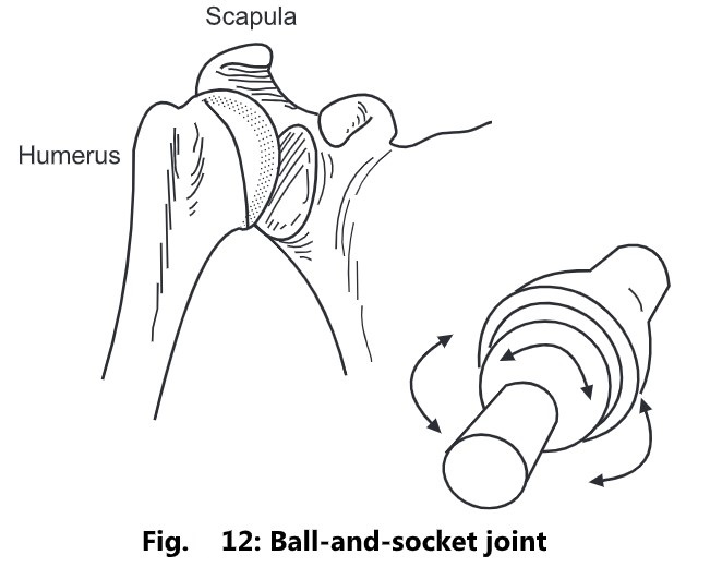
Table 2: Different types of Synovial Joints